GIDEON blog

Free Demo ► Latest Updates ► Video Tutorials ►
Subscribe , j x

Visceral Leishmaniasis in Spain

Although leishmaniasis has generated increasing concern in Spain during recent years, the incidence of reported cases has changed little during the past 25 years. The following data are abstracted from the GIDEON e-book series [1,2].

Time and Place:
Visceral leishmaniasis is most common in the southern region.
– Seroprevalence among dogs in this region is lowest during April, peaking during October.
– A restricted focus has been identified in the Piorat region of northeastern Spain.
– A single autochthonous case has been reported from Minorca.
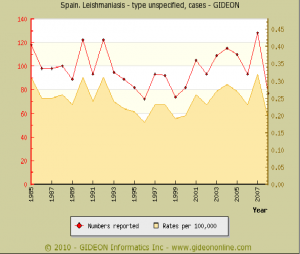
Graph Notes:
1. 1,180 hospitalizations were reported for leishmaniasis during 1999 to 2003 (0.4 per 100,000 population).
2. 11 cases (0 fatal) were identified among immunocompetent children in southern Madrid during 1994 to 2007.
3. 60 deaths were ascribed to leishmaniasis during 1980 to 1994.
4. In 2005, three cases were reported among Romanian workers returning from Spain.

Leishmaniasis and HIV infection:
– 450 AIDS/Leishmania coinfections were reported nationwide during 1990 to 1995 (52.4% of the world’s total coinfections); 412 during 1996 to 1998; 1,099 as of 2001.
– 835 cases of HIV-Leishmania coinfection were reported during January 1990 to June 1998; 412 during January 1996 to June 1998; 1,099 during January 1990 to January 2001; 130 during January 2001 to December 2006.
– Spain accounted for 58% of all such coinfections in southwestern Europe during 1990 to 1998.
– 2.49% of AIDS patients in the Madrid region are coinfected with visceral leishmaniasis.
– 228 episodes of visceral leishmaniasis were diagnosed in 155 HIV-infected patients in Valencia and Murcia during 1988 to 2001.

Transmission through sharing of needles by injecting drug abusers has been reported.
– Approximately 30% of IVDA in southern Spain are seropositive.
– Evidence for Leishmania is found in 52% of discarded syringes collected in southern Madrid (1998) and 34% from southwestern Madrid (2000 to 2001).

Leishmania infantum has been identified in some cases of cutaneous and mucosal leishmaniasis.

Prevalence surveys:
2.4% of asymptomatic blood donors in Elvissa (Belearic Islands) have been found to have cryptic infection by ELISA – 7.6% by Western blot (2004 publication)
20.5% of wolves (Canis lupus), 14.1% of foxes (Vulpes vulpes), 28.6% of Egyptian mongooses (Herpestes ichneumon), 1 of 4 genets (Geneta geneta), and 1 of 4 Iberian lynxes (Lynx pardinus) (2008 publication)
29.4% of dogs in the Barcelona region (PCR, 2009 publication)
19.6% of seronegative blood samples in a canine blood bank. (Leishmanial DNA, Barcelona, 2008 publication)

Seroprevalence surveys:
3.1% of blood donors from the Balearic Islands. (Majorca, Formentera, and Minorca, 2008 publication)
4.9% of the general population in Castilla Leon, and 64.0% of HIV-positive patients (1996)
11.5% of children and 52.8% of adults in Alicante, southeastern region are skin test-positive (1997 to 1998)
5.25% of dogs in the Madrid area
7.8% of stray dogs in the Madrid region (1996 to 2006)
13% of dogs on Mallorca are infected; 3.7% in northwestern Spain (2004 publication)
30% of dogs in northeastern Spain (2006 publication)
22% of dogs in Crevillente town, Alicante (1999)
13.0% of dogs in the Alpujarras region of southeastern Spain – with highest rates in larger, older dogs which live outdoors (2006)
8.1% of dogs in the Madrid region (2006 to 2007)
5.3% of shelter dogs in central Spain (2010 publication)
3% of cats in Barcelona (2007 publication)
1.29% of cats in Madrid (2005 to 2006)
6.29% of cats in the northeastern Iberian Peninsula and Balearic Islands (2007 publication)

Leishmania infantum has been identified in captive wolves (Canis lupus) in northern Spain (2008 publication)

Vectors:
The vectors in Spain are Phlebotomus (Larroussius) ariasi and Ph. (La.) perniciosus.

References:
1. Berger SA. Infectious Diseases of Spain. https://www.gideononline.com/ebooks/country/infectious-diseases-of-spain/ , 516 pp. 2010, Gideon Informatics, Inc.
2. Berger SA. Visceral Leishmaniasis: Global Status. https://www.gideononline.com/ebooks/disease/visceral-leishmaniasis-global-status/ , 84 pp. 2010, Gideon Informatics, Inc.
Malaria in Ireland

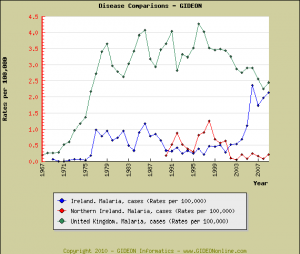
Although increasing numbers of malaria cases have been reported in Ireland in recent years, disease rates for Northern Ireland and the United Kingdom as a whole have actually declined. [1,2]

References:
1. Berger SA. Infectious Diseases of Ireland and Northern Ireland, 2010. Gideon e-book series, 437 pp. https://www.gideononline.com/ebooks/country/infectious-diseases-of-ireland-and-northern-ireland/
2. Berger SA. Malaria: Global Status, 2010. Gideon e-boon series, 378 pp. https://www.gideononline.com/ebooks/disease/malaria-global-status/

Tick-borne Diseases of Russia

Regarding recent TBE activity in Altai and Omsk, a total of 26 human zoonoses are transmitted by ticks. Thirteen of these occur in Russia: [1]

Anaplasmosis
Astrakhan fever
Babesiosis
Crimean-Congo hemorrhagic fever
Japanese spotted fever
Lyme disease
North Asian tick typhus
Omsk hemorrhagic fever
Powassan
Relapsing fever
Rickettsial Spotted fevers (ie, Rickettsia slovaca infection)
Tick-borne encephalitis
Tularemia

References:
1. Berger SA. Infectious Diseases of the Russian Federation. 2010, Gideon e-book series, 461 pp. https://www.gideononline.com/ebooks/country/infectious-diseases-of-the-russian-federation/

Dengue in Egypt

The following backround data on dengue in Egypt are abstracted from the GIDEON e-book series. [1,2]

Historical background:
– Outbreaks of dengue were reported in Cairo and Alexandria during 1799; and in Port Said in 1871.
– Subsequent cases were reported in Cairo and the sea ports in 1880; and in the Nile delta in 1889.
– Dengue fever was widespread during the first half of the 20th century.
– A decline in transmission was recorded in Egypt after 1940, and was ascribed to rapid decrease of Stegomyia (Aedes) aegypti populations with the introduction of DDT during and after the Second World War.

Notable outbreaks:
1928 – An outbreak (70 fatal cases) was reported.
1937 – An outbreak (50 fatal cases) was reported.

References:
1. Berger SA. Infectious Diseases of Egypt. 2010, Gideon Informatics, Inc., 367 pp. https://www.gideononline.com/ebooks/country/infectious-diseases-of-egypt/
2. Berger SA. Dengue: Global Status. 2010, Gideon Informatics, Inc., 191 pp. https://www.gideononline.com/ebooks/disease/dengue-global-status/

Eosinophilic Meningitis in Australia

Recently, a man in New South Wales acquired eosinophilic meningitis (angiostrongyliasis) after ingesting a slug.

The 1st Australian case of human angiostrongyliasis was reported in 1971, in Brisbane.

At first, sporadic cases were reported only from coastal Queensland, northern New South Wales.

Four cases were reported in Victoria to 1999 (3 of these imported from Fiji; one fatal case).

Seropositivity is common among aboriginals.

Reservoirs:
The parasite has been identified in dogs in Sydney as well as in grey-headed fruit bats (Pteropus poliocephalus) Bennett’s wallaby (Macropus rufogriseus) rufous bettongs (Aepyprymnus rufescens) and captive tamarins (Sanguinus spp).

Further information regarding this, and all other diseases endemic to Australia, is available in Infectious Diseases of Australia. Los Angeles, Gideon Informatics, 2010. 488 pp. [edited] www.gideononline.com/ebooks/country/infectious-diseases-of-australia/

Update: Published in ProMED

Kunjin Virus

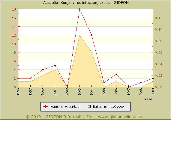
Kunjin virus (KUN) is similar Murray Valley encephalitis (MVE) virus; however, infection by KUN is generally milder than that with MVE, with a lower rate of encephalitis. KUN appears to have been responsible for some of the ‘Murray Valley encephalitis’ cases reported in 1974, and was implicated in an additional sporadic case in northern Victoria in 1984. KUN-is more widely distributed than MVE, and is found in most of tropical Australia, eastern Queensland, and occasionally southeastern Australia.

Notes for individual years:
1996 – Two cases in Queensland.
1997 – Two cases in Western Australia and two in Northern Territory. A presumptive case of KUN was reported in Pilbara, WA
2000 – Three in Western Australia and one in Northern Territory.
2001 – New South Wales, Northern Territory [2 cases] and Western Australia.
2003 – All cases reported from Queensland
2004 – 11 in Queensland and 1 in Victoria.
2005 – 1 in Queensland
2006 – 2 in Western Australia and 1 in Queensland

The vectors for KUN are Culex annulirostris, Cu. pseudovishnui and Cu. squamosis.

An extensive review of Australo-Pacific Arbovirus diseases is available in the ebook Australo-Pacific Arboviruses: Global Status

Update: posted in ProMED

Leptospirosis in Argentina

Leptospirosis was first identified in Argentina in 1915.
– Most cases are reported from Greater Buenos Aires.
– 276 cases were diagnosed in the Greater Buenos Aires area during 1990 to 1999 – 43 of these characterized by pneumonia.
– 32 cases (6 fatal) were reported in Santa Fe during the first two months of 2010.

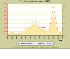
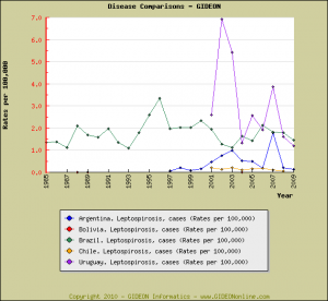
The following graphs depict incidence and case rates of Leptospirosis in Argentina, and contrast these data with those of surrounding countries:


Notes:
Individual years:
1998 – Included 53 cases in Santa Fe and 16 in Buenos Aires.
2001 – Included 63 cases in Santa Fe and 89 in Buenos Aires.
2005 – Three fatal.

Twelve serovars were identified in human infection during the 1970’s, with most cases due to serovars bratislava, icterohaemorrhagiae and butembo.
– 41% of infections during 1984 to 1994 were due to Leptospira interrogans canicola.

Seroprevalence surveys:
15.7% of the population, nationwide (1976 publication)
8.7% of healthy persons in Corrientes and 0.5% in Neuquen are seropositive (1979 publication)

Reservoirs:
The principal hosts in this country are rats, pigs and dogs.
– 25% to 40% of rats and 10% to 60% of dogs in Buenos Aires are seropositive – most often toward L. interrogans canicola and L. i. pyrogenes.
– Antibody toward serovars pomona, hebdomadis group, pyrogenes, tarassovi, and canicola has been demonstrated in healthy horses.

Notable outbreaks:
1976 – An outbreak (10 cases) in Buenos Aires followed swimming in a drainage canal contaminated by pigs.
1977 (publication year) – An outbreak was reported among children in Longchamps, Buenos Aires Province.
1998 – An outbreak (12 confirmed and 2 suspicious cases) in Santa Fe was related to water.
2004 – An outbreak (12 suspect cases, 1 fatal) was reported in Entre Rios.
2007 – An outbreak (400 cases or more) was reported in Santa Fe.

Vaccination Coverage in Haiti

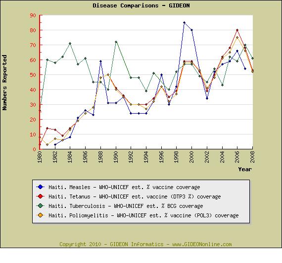
Although vaccination coverage for major infectious diseases in Haiti was below that of other countries in the region, recent WHO estimates had shown some improvement in recent years. Most recent data are summarized in the following chart:

Also check out GIDEON’s free ebook: Infectious Diseases of Haiti

Leptospirosis in Queensland

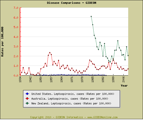
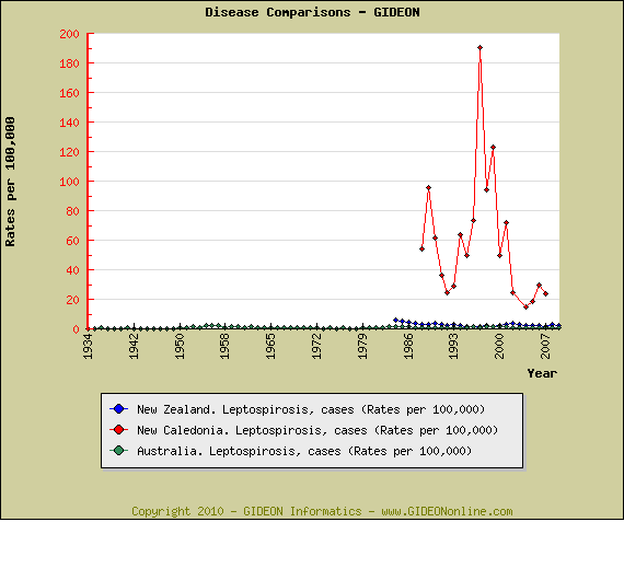
A recent ProMED warning to backpackers is well taken – leptospirosis is a common disease in the Pacific region. In the following graphs I’ve compared disease rates for Australia and New Zealand with those of the United States.

Leptospirosis rates in nearby New Caledonia are even more striking, and exceed those of salmonellosis, hepatitis A, hepatitis B, gonorrhea, and tuberculosis.

Updated Microbiology module

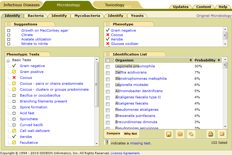
GIDEON’s redesigned Microbiology module has been launched (screenshot). Following the update of the GIDEON Diagnosis module, we’ve implemented many of the new features in Microbiology, including

  • New tabs
  • Suggestions
  • Dynamic identification
  • Usability improvements

New tabs

The new tabs in Microbiology replace the older radio buttons and make it easier to reach each function. For each category: Bacteria, Mycobacteria and Yeasts there is easy access to Identify or Lists.

Suggestions

GIDEON’s Microbiology Compare function, until now, has ranked phenotypic tests which are most likely to impact the Identification list. Now, the top four tests which are most likely to focus and shorten the list of possible organisms are displayed and dynamically updated as each new test result is entered. Clickable boxes which allow the user to enter a “yes”, “no” or “unknown”, appear and enlarge each time the mouse passes near a test.

Dynamic identification

The Identify button has been eliminated! Now, the Identification list is automatically updated as test results are entered. This feature demonstrates the effects of each new test result as it is entered.

Diagnosis results buttonsThe familiar buttons: Compare, Why Not, Open case, Save case, Remove All, Print, Email are all in the Identification list area.
Usability improvements

Updated lists

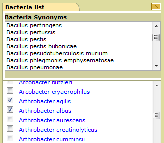
The organism lists have been updated to extend the length of the screen. The organisms can be selected by clicking the check boxes to their left and then Compared. Clicking on expands the list of synonyms.

Collapsible windows

Windows in Identify, such as Suggestions and Phenotypic Tests can be minimized and hidden. For example to not see suggestions, click on the minimize button Minimize button to the left title.

Mouse overs
More mouse-overs have been added: Clickable boxes expand as you mouse over them, and display clear symbols to select “yes” or “no.”

Phenotype
You can now click on the tests  in the Phenotype window.

Quick sorting
Probability sort arrowIdentification results can be sorted alphabetically or by probability easily by clicking the column title.
Window resizing
Changing vertical window size expands size of Phenotypic tests and Identification list sub-windows in Identify and Organism list. This is a great feature for larger monitors.

Previous version
Click “Original Microbiology” to use the older interface.

Update: See video demonstration

GIDEON Sign In