GIDEON blog

Free Demo ► Latest Updates ► Video Tutorials ►
Subscribe , j x

Archive for the ‘Epidemiology’ Category

Less Prep, More Insights: GIDEON R for Epidemiology-Related Research

We are excited to introduce the GIDEON R Package, released this month as a beta test for our researchers worldwide. GIDEON R is an efficient plug-and-play statistical tool for all researchers to clean, analyze, and visualize their epidemiological data from the GIDEON database. There is no need to program your own REST API queries. 
In the past 3 years, 200+ research papers used GIDEON data

EXCLUSIVE SNEAK PEEK FOR EXISTING GIDEON CUSTOMERS

  • All existing GIDEON customers get early access to experience and test the GIDEON R Beta package. Click here to download the package.
  • Let us know what you think! Email us at kristina@gideononline.com and tell us what you liked and what could be improved.
  • Your comments help us refine and launch the best version of GIDEON R for your research needs.
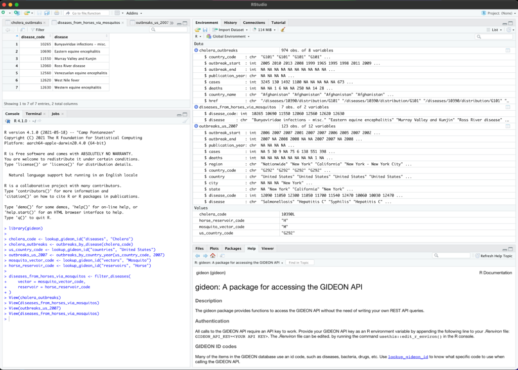
GIDEON R package in use
Image: GIDEON R package in use


Over 200 scientific studies that leveraged GIDEON’s (Global Infectious Diseases and Epidemiology Network) database were published in just the past three years. This number is rapidly growing as many researchers turn to the extensive infectious disease database for epidemiological insights and cross-discipline studies. 

The GIDEON database is a valuable web-based reference of infectious diseases and their global occurrences since the 1900s. As of 2020, researchers also get to customize and build their statistical tools to analyze data using the GIDEON API or Application Programming Interface (API). 

While GIDEON’s massive global database of infectious diseases and the GIDEON API are potent tools, GIDEON R allows you to boost your analytics to the next level. 

Why use GIDEON R?

R, the free, open-source programming language, has transformed how researchers prep, clean, and wrangle large databases to get good information. With minimal coding required, researchers can clean and set up their data faster and conduct reproducible steps during data analysis. 

R is quickly gaining in popularity with researchers. R programming is a part of the standard data analytics curriculum in universities like Harvard and the Imperial College in London. It is fast becoming a mainstay in Public Health and epidemiological research studies and reports. In the UK, the O Health foundation, an independent charity, developed an NHS-R community to help leverage the power of R for the NHS (National Health System). Teams of experts trained NHS analysts to use and embed R into the NHS to help improve the delivery of care.  

Using the GIDEON R package brings you more efficiency to:

  • Investigate 25,000+ ongoing and historical infectious disease outbreaks,
  • Produce granular outbreak maps for chosen diseases in a given year range
  • Study the emergence of zoonotic diseases in a particular country,   
  • Evaluate epidemiological situations around the globe, 
  • Retrieve a wealth of information on 360+ infectious diseases, 2,000+ pathogens, and 30,000+ trade names of drugs and vaccines.

The GIDEON R package brings all the convenience and efficiency of the free, open-source programming language R to the world of epidemiological research. 

Benefits of using GIDEON R include…

  • Simplicity: 

There is no need to learn how to work with a REST API client to parse the GIDEON database. With GIDEON R, you can hit the ground running and start crunching your data. 

Working with a GIDEON REST API offers you greater and complete control over how your program manipulates data. However, it requires users to possess programming skills and may also mean an investment in rigorous manual and automatic testing to ensure it functions well under pressure. Your team will need to spend considerable time testing, sequencing API calls correctly, validating parameters, and fixing any other issues before beginning the analysis. GIDEON R gives researchers familiar with R the ability to skip this part of the process and get straight to the analytics.  

  • Reproducibility: 

GIDEON R allows you to create scripts for your entire data analysis process and run a simulation. This way, even if you make a mild edit to the data, the whole process can be run again with the reassurance of reproducibility. As a researcher, you can then focus on developing and analyzing different runs without worrying about the analytical method changing. 

  • Flexibility

Epidemiological research is complex and challenging. No two studies will ever be precisely the same. R offers a considerable toolkit of statistical modeling tools that epidemiologists require, including logistic and Poisson regression and Cox proportional hazard models. 

  • Better Visualization

With R, data comes to life. Using R for data visualization is like the famous scene in the classic movie ‘The Wizard of Oz’ when Dorothy steps out of her dull black and white house and into the dazzling technicolor land of Oz. 

R can create any type of graph or charts – fast ones for analysis and even publication-ready charts with minimum code. R offers in-built functions and libraries to generate basic maps like bar charts, histograms, and scatter plots. It can also create advanced visualization tools like heat and mosaic maps, 3D graphs, or correlograms in vivid technicolor for your exploratory data analysis, presentations, and publications.

  • Compatibility

R runs on everything. R’s code is platform-independent – which means it does not matter if you use Windows, Mac, or any other system. So, with GIDEON R, you can be sure that your program is compatible with any type of platform you or your team use. This is a significant benefit when working with teams located in different regions and across the globe. 

GIDEON R optimizes how researchers use the GIDEON API to mine the GIDEON infectious disease database for epidemiological research. 


Want to be one of the first to try the new GIDEON R package?

  • If you are an existing GIDEON customer, click here to sign up for our beta test. Please give us your feedback at kristina@gideononline.com.
  • If you are not an existing GIDEON customer but would like to be, sign up for a free demo to get started.


GIDEON API

The GIDEON API allows medical professionals and researchers pressed for time and resources access to global data on hundreds of diseases, drugs, and bacteria – since 1348 AD. 

With the GIDEON API, you get a direct feed of infectious disease data from around the world at your fingertips. The GIDEON API is based on RESTful principles, and data is refreshed and updated every day, sometimes even multiple times a day. 

The best part? All institutional subscribers to GIDEON get access to the GIDEON API free of charge. 


Published articles that used GIDEON

Epidemic infectious disease outbreak with person analyzing virus strain and worldwide situation. SARS-CoV-2 pathogen causing coronavirus covid-19 pandemic disrupting social and economic life

According to Professor Rodolphe Desbordes, Professor of Economics at SKEMA Business School, France, and widely published in International Economics and Economic Development: 

“GIDEON was the perfect database for the epidemiological project I had in mind <…> the information provided on each disease was crucial to a better understanding of disease-specific characteristics.“


GIDEON has a rich history of partnering with researchers and scientists worldwide by offering a wide variety of resources on infectious diseases. You can find data going back to 1348 AD, track outbreaks on an interactive map, identify over 2000+ pathogens, diagnose and compare any number of infectious diseases, drugs, and microbes. 

The GIDEON database contains 23,600 country-specific notes with 3+ million words of text that outline the status of specific infections within each country. Also featured are over 250,000 linked references, 3,000 images, 34,000 graphs, and numerous interactive maps. 

There are more than 200 studies published in just the past three years that use GIDEON’s database to generate meaningful insights. Here are a few of the recent articles published that used GIDEON for their research: 

  • June 2021, Dengue: Alisa Aliaga-Samanez et al. from Spain published the first high-resolution analysis of biogeographic changes in dengue transmission risk. The study informs about the Dengue virus (DENV) making a home in previously low-risk areas and urges the global public health community to implement preventive measures [1]. 


  • June 2021, Foodborne Parasitic Diseases: F. Chavez-Ruvalcaba et al. published their review of foodborne parasitic diseases in the neotropics. Since more than one-fifth of the world’s population is infected by one or more intestinal parasites, the authors review the most common ones affecting countries in Central and South America [2]. 


  • June 2021, Lyme Borreliosis in Poland: Brzozowska et al. published their study about the tick-borne Lyme Borreliosis in Poland. They found the incidence to be equally significant in urban and rural communities and stressed the importance of widespread awareness and education. The study used GIDEON-generated data to compare Lyme Borreliosis prevalence across the globe [3].  


  • May 2021, Control of Intestinal Nematodes in African Green Monkeys (AGMs): A veterinary study by Katalina Cruz et al. tackled the efficacy of antiparasitic treatment and husbandry methods to control nematode infections in AGMs. The authors referred to insights from the GIDEON database to highlight that because AGMs regularly come in contact with humans on the island, they may play a role in the zoonotic parasitic infections commonly found on St. Kitts [4]. 


  • May 2021, Emerging Antibiotic-Resistant Pathogens in Iran: As part of their study, Rahder et al. analyzed the reported prevalence of actinomycetes infections worldwide using published global prevalence data sourced from the GIDEON database. They identify infections in Iran affecting immunocompromised and other vulnerable patients and recommend continuous monitoring to better prevent infection and improve therapeutic methods to treat the infections [5]. 


  • March 2021, Brucellosis: Battikh et al. from Tunisia used the GIDEON database to analyze the rise in Brucellosis cases in their hospital. They found that osteoarticular involvement was the most common complication of brucellosis in their patient pool. The researchers recommended better animal control practices through vaccinations, occupational and personal hygiene, farm sanitation, and more to lower the number of cases [6]. 


  • March 2021, Global Empirical Assessment of Spatial Dynamics of Major Disease Outbreaks: Professor Rodolphe Desbordes presented the spatio-temporal dependence and mortality consequences of the top 15 disease outbreaks in developed or developing countries over ten years. In the article, he states that his team mainly relied on the “under-exploited GIDEON database that provides a worldwide coverage of all infectious diseases [7].”

    In an interview, Professor Desbordes talked about why having access to data added to his study. He mentioned, “As an applied economist, I value excellent data on a novel and interesting issue more than anything else. The GIDEON database allowed me to publish in an excellent journal and, most importantly, carefully model the spatial diffusion of infectious diseases in a globalized world.”


  • March 2021, Ecological Conditions That Increase Vector-Borne and Zoonotic Diseases Outbreaks: Morand and Lajaunie published their findings on how global forest cover changes and oil palm expansions are associated with increased outbreaks of vector-borne and zoonotic disease outbreaks from 1990 – 2016. The authors state, “Here, we examine the global trends between changes in forest cover in recent decades and epidemics of human infectious diseases, using the GIDEON global database, which is the best available dataset on infectious diseases that has already been used in several studies [8].”


Want to be an early user and test GIDEON R?

  • All existing GIDEON customers get free access to experience and test the GIDEON R Beta package. Click here to start.
  • Let us know what you think! Email us at kristina@gideononline.com with what you liked and what could be improved.
  • Not an existing GIDEON customer? Don’t worry. We’ve got you covered. Sign up here for a free demo to get started.

Conclusion

GIDEON is one of the most well-known and comprehensive global databases for infectious diseases. Data is refreshed daily, and the GIDEON API allows medical professionals and researchers access to a continuous stream of data. 

The GIDEON R package allows researchers to retrieve, clean, analyze, and visualize infectious disease data in real-time from the GIDEON database without the need to get familiar with API clients and learn to program your own API queries. This improves the efficiency and reproducibility of research methods and results and lowers the time and costs required to learn how to work with REST APIs.


Did you like this article? Please share it on social media!

Check out more of our latest content here


References

[1]  A. A.-S. e. al., “Worldwide dynamic biogeography of zoonotic and anthroponotic dengue,” PLoS Negl. Trop. Dis., vol. 15, no. 6, p. e0009496, 2021. 
[2]  F. Chávez-Ruvalcaba, M. I. Chávez-Ruvalcaba, M. K. Santibañez, J. L. Muñoz-Carrillo, C. A. León and R. R. Martínez, “Foodborne Parasitic Diseases in the Neotropics – a review,” Helminthologia, vol. 58, no. 2, pp. 119-133, 2021. 
[3]  M. Brzozowska, A. Wierzba, A. Śliwczyński, M. Myśliwiec, K. Kozłowski and W. Wierzba, “The problem of Lyme borreliosis infections in urban and rural residents in Poland, based on National Health Fund data,” Annals of Agricultural and Environmental Medicine, vol. 28, no. 2, p. 277–282, 2021. 
[4]  K. Cruz, T. M. Corey, M. Vandenplas, M. Trelis, A. Osuna and P. J. Kelly, “Case report: Control of intestinal nematodes in captiveChlorocebus sabaeus,” Onderstepoort Journal of Veterinary Research, vol. 88, no. 1, pp. 2219-0635, 2021. 
[5]  H. A. Rahdar, S. Mahmoudi, A. Bahador, F. Ghiasvand, H. Sadeghpour and M. M. Feizabadi, “Molecular identification and antibiotic resistance pattern of actinomycetes isolates among immunocompromised patients in Iran, emerging of new infections,” Scientific Reports, vol. 11, 2021. 
[6]  H. Battikh, A. Berriche, R. Zayoud, L. Ammari, R. Abdelmalek, B. Kilani, H. Tiouiri Ben Aissa and M. Zribi, “Clinical and laboratory features of brucellosis in a university hospital in Tunisia,” Infectious Diseases Now, 2021. 
[7]  R. Desbordes, “Spatial dynamics of major infectious diseases outbreaks: A global empirical assessment,” Journal of Mathematical Economics, vol. 93, 2021. 
[8]  S. Morand and C. Lajaunie, “Outbreaks of Vector-Borne and Zoonotic Diseases Are Associated With Changes in Forest Cover and Oil Palm Expansion at Global Scale,” Front. Vet. Sci., vol. 8, p. 230, 2021. 
[9]  R. e. al., “Data proliferation, reconciliation, and synthesis in viral ecology,” bioRxiv, 2021. 

New Dengue Study Identifies New High-Risk Countries

Pathogen of the month: Dengue virus


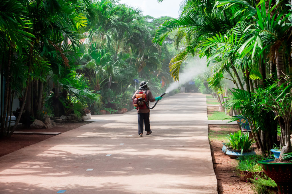
A man using a sprinkler ULV type for kill mosquito carrier of Zika virus and dengue fever it outbreak in school at the rainy season.
Image: Sprinkling to kill Aedes aegypti, a vector of Dengue, during the rainy season.


written by Chandana Balasubramanian

Halfway through every Hollywood disaster movie, when all hell breaks loose, a few rogue scientists are brought in. This handful of experts would have predicted the disease outbreak or catastrophe years, or sometimes decades, before. But their advice would have fallen on deaf ears – until it was almost too late.

Meanwhile, in real life, a large and vocal group of researchers and public health organizations are sounding the alarm about Dengue as a pandemic-level threat in the near future.

The relatively good news is that Dengue, a vector-borne illness, is not transmissible between humans. So, we can prevent a full-blown Dengue pandemic if the global community of public health officials:

But why is Dengue such a colossal threat? Why is prevention important?


Dengue cases on the rise

Currently, Dengue cases are estimated to be anywhere from 5 – 100 million. This wide range is because many cases stay unreported, or the symptoms of Dengue are confused for other diseases. But the number of Dengue cases is expected to boom in the next few decades, with 60% growth by 2080 [1].

The latest study on the spread of Dengue is by Aliaga-Samanez et al. from Spain [2]. The study is the first high-resolution analysis of how the risk of Dengue transmission has been changing geographically since the late 20th century. The study uses robust databases like the World Health Organization (WHO) [1], and the Global Infectious Disease and Epidemiology Online Network (GIDEON) [3] to track and map global Dengue cases since the 1900s.

All Dengue experts agree that the number of Dengue cases is rising fast and spreading wide across the globe. WHO estimates that about half the world’s population is now at risk [4]. Aliaga-Samanez et al. identified that the Dengue virus (DENV) has been making a home in previously low-risk areas, potentially due to global warming and deforestation. The authors also report that while the Aedes mosquito is responsible for Dengue transmission, DENV can be spread by primates and could adapt to be transmissible by other vectors.

Tracking the geographical spread of the Dengue virus by different vectors is complex. According to the primary researcher Alisa Samanez, using the GIDEON database – “one of the most complete data sources worldwide on zoonoses” – to build their own database was a significant factor to track a zoonotic disease like Dengue.


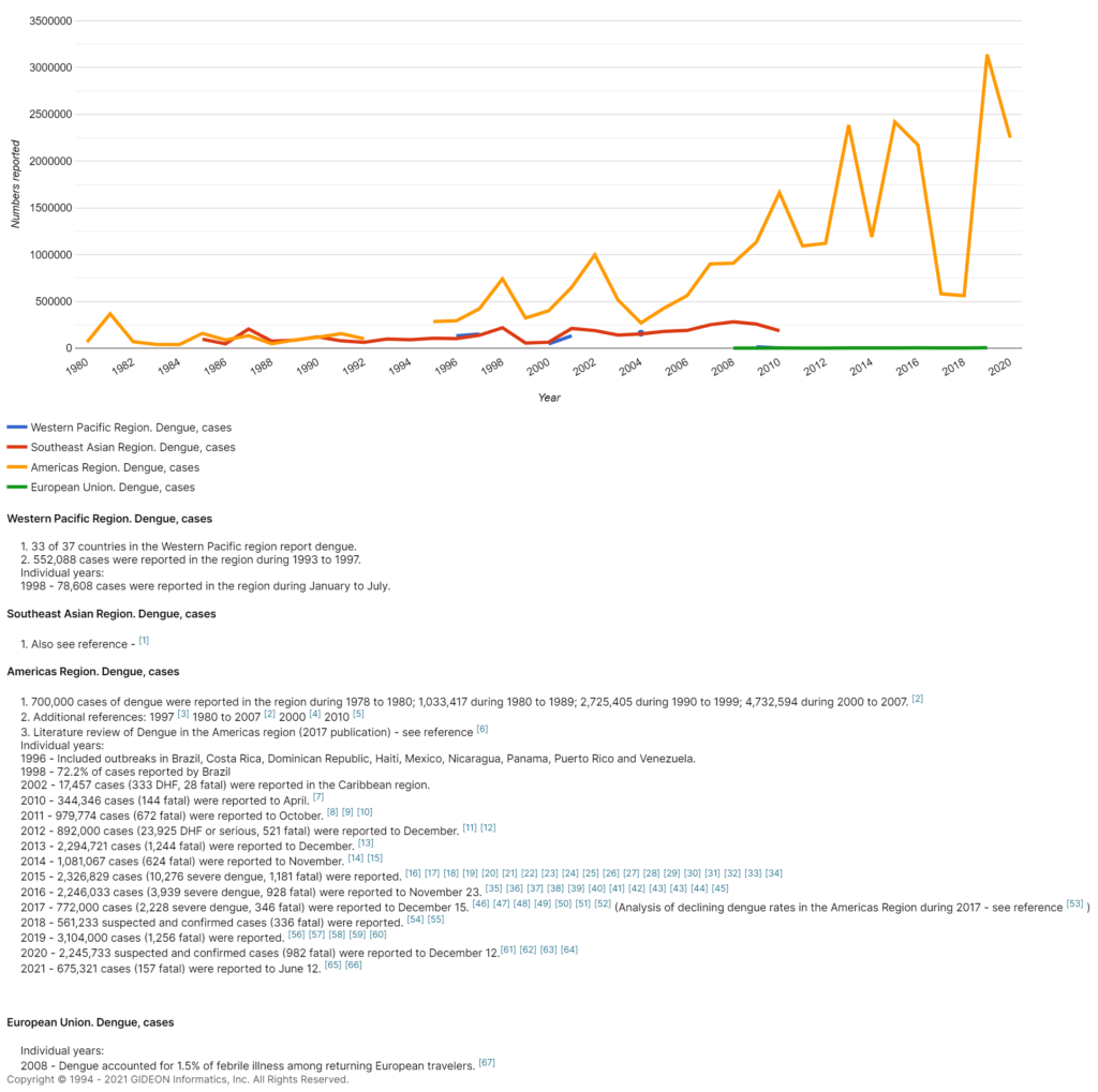
Reported Dengue cases in different regions, 1980 – 2020

Graph illustrating Dengue cases in different regions, 1980 - 2020
Image: Graph illustrating Dengue cases in different regions, 1980 – 2020. Copyright © GIDEON Informatics, Inc.


Dengue mosquitoes spread their wings worldwide

While Dengue cases are primarily found in the tropical regions of Asia, Africa, and the Americas, this is rapidly changing. The study from Spain determined that other regions now at risk are South-East China, Papua New Guinea, North Australia, South USA, parts of Colombia, Venezuela, Madagascar, and even Japan and South and Central Europe. The study predicts that Dengue could spread to Argentina and South-West Asia, from Pakistan and the Arabian Peninsula.

Ideally, public health officials in these regions will begin training their healthcare professionals to diagnose and treat Dengue early and raise awareness among their populations.  

For example, initially, Egypt’s Ministry of Health dismissed reports of Dengue fever in certain regions. But eventually, they reviewed published reports of Dengue outbreaks in studies that used GIDEON’s clinical tools. This prompted them to develop a training program for their health workers for the early detection and treatment of Dengue [5].

Two significant factors accelerating the spread of Dengue worldwide are the 2.9 trillion U.S dollar global travel and tourism industry and climate change.

Many studies indicate that international travelers are at considerable risk for Dengue spread. Ratnam et al. from Australia concluded that Dengue infections in international travelers occur frequently and may be associated with substantial morbidity [6]. 

What is worrisome is that while Dengue is not directly contagious between humans, it is transmittable from an infected human to an infection-free Aedes mosquito. So, if a Dengue-infected individual travels to another country during the viremic or infection-spreading period, a native Aedes mosquito may bite the individual and become infected [7]. This newly infected mosquito can infect other individuals.

Additionally, climate change is a contributor because rising temperatures in previously colder environments are fertile grounds for mosquitoes to thrive. Higher temperatures also shorten the cycle of a mosquito picking up a Dengue infection and transmitting it.


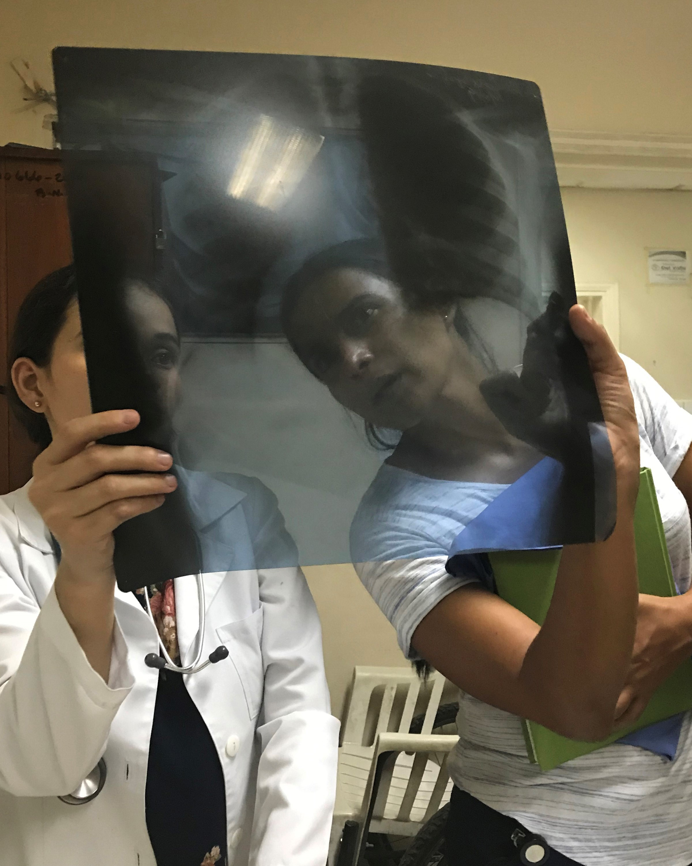
Captured in La Paz, Honduras, this August 2019 photograph, depicted Dr. Liliana Sanchez-Gonzales on the left, an Epidemiologist with the Centers for Disease Control and Prevention’s (CDC) Dengue Branch, along with an unidentified Honduran physician, as they were examining the chest x-ray of a patient, who was on the verge of developing severe dengue. The x-ray revealed the presence of fluid in her lungs, possibly due to plasma leakage, as she was going into dengue-related shock.
Image: Captured in La Paz, Honduras, this August 2019 photograph, depicted Dr. Liliana Sanchez-Gonzales on the left, an Epidemiologist with the Centers for Disease Control and Prevention’s (CDC) Dengue Branch, along with an unidentified Honduran physician, as they were examining the chest x-ray of a patient, who was on the verge of developing severe dengue. The x-ray revealed the presence of fluid in her lungs, possibly due to plasma leakage, as she was going into dengue-related shock.


Why is Dengue dangerous?

According to WHO, severe Dengue is a leading cause of serious illness, hospitalization, and death among children and adults in some Asian and Latin American countries [4]. Severe Dengue involves severe bleeding, liver, heart, and other organ impairment, and plasma leakage.

Many Dengue infections are mild with flu-like symptoms, but a lack of awareness and early detection could lead to severe Dengue. And since the incubation period varies from four to ten days, it may be overlooked or misdiagnosed until it becomes severe. Additionally, because there are four different types of the Dengue virus, individuals once affected by Dengue can be re-infected up to four times.

The Dengue vaccine is only available in certain countries and, as per WHO, is restricted to those aged 9 to 45 and individuals previously infected by DENV. Hopefully, we get a better alternative. But until then, we need to raise awareness about early detection and treatment on the frontlines and track outbreaks closely.


Conclusion

If we have learned anything from the COVID-19 pandemic, it is this: the best disaster control is early detection and prevention. According to Aliaga-Samanez et al., Dengue is poised to be the next pandemic. Only through global collaboration, rigorous tracking, and preventive public health programs can we banish health catastrophes like a Dengue pandemic to the world of fiction – away from our collective reality.


Did you like this article? Please share it on social media!

Check out more of our latest content here


References

[1] World Health Organization, “Global strategy for Dengue prevention and control, 2012-2020,” World Health Organization, Genève, Switzerland, 2012.
[2] A. A.-S. e. al., “Worldwide dynamic biogeography of zoonotic and anthroponotic Dengue,” PLoS Negl. Trop. Dis., vol. 15, no. 6, p. e0009496, 2021.
[3] B. S, “Dengue: Global Status,” GIDEON Informatics, Inc., Los Angeles, California, USA, 2015.
[4] W. W. H. Organization, “Dengue and Severe Dengue Fact Sheet,” World Health Organization, 19 May 2021. [Online]. Available: https://www.who.int/news-room/fact-sheets/detail/Dengue-and-severe-Dengue. [Accessed 04 07 2021].
[5] Department of Tropical Medicine, Ain Shams University, Cairo, Egypt, “Dengue fever, Correspondence to Nadia A Abdelkader, MD,” Egypt J Intern Med, vol. 30, pp. 47-48, 2018.
[6] F. Irani Ratnam, “Dengue Fever and International Travel,” Journal of Travel Medicine, vol. 20, no. 6, p. 384–393, 2013.
[7] J. P. Messina, “The current and future global distribution and population at risk of Dengue,” Nature Microbiology, vol. 4, p. 1508–1515, 2019.

Monkeypox Virus in Texas: Are We Ready to Prevent the Next Outbreak?

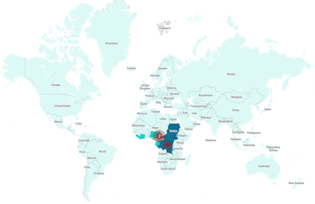
Monkeypox outbreaks map 2018-2021
Image: World map of Monkeypox outbreaks, 2018-2021. Copyright © GIDEON Informatics, Inc.


written by Chandana Balasubramanian

Hot on the heels of the COVID-19 virus and its variants, another infectious virus recently landed in the United States. On July 9th, 2021, a passenger with monkeypox flew from Lagos in Nigeria to the United States, ultimately landing in Dallas, Texas. The Centers for Disease Control and Prevention, CDC confirmed the case of Monkeypox on July 15th – the first case of monkeypox in the United States in almost 20 years. While rare, the Monkeypox virus is contagious and kills one in ten people that it infects.


“…we are only a plane ride away from any global infectious disease.”

– Dr. Philip Huang, Director of the Dallas County Health and Human Services.


A few fortunate events prevented a Monkeypox outbreak in the United States this time around. 

  1. Symptoms Presented Early: Symptoms of monkeypox are similar to those of smallpox and can take anywhere from 7 to 14 days to appear after getting infected (incubation period). Luckily, symptoms presented earlier, and the patient visited a hospital within four days of entry into the country. 
  2. Early Detection by Frontline Clinicians: The symptoms of monkeypox include fever, headache, backache, swollen lymph nodes, chills, and fatigue. Within three days of the onset of fever, a rash appears on the face and spreads to other parts of the body. Doctors and healthcare workers at the Dallas hospital were able to identify and diagnose monkeypox quickly, so the infected patient was isolated early. 
  3. Mask Mandates and Social Distancing: Monkeypox can spread from person to person through respiratory droplets and body fluids. In this instance, a huge silver lining is that due to COVID-19 mandates, everyone, including the patient, other passengers, airline and airport staff, and others wore masks. This helped prevent the spread. 

Monkeypox is transmitted to humans through infected animals or other humans. Animals can share it through bites, scratches, or direct contact with body fluids. Human transmission is mostly through respiratory droplets and body fluids but also contact with contaminated clothes. 

While there was no outbreak this time, there have been a few in prior years.

***

Monkeypox Outbreaks

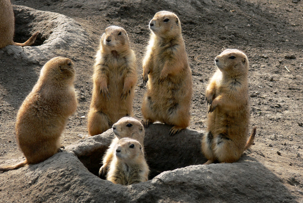
In the case of monkeypox, there have been notable outbreaks in the past. In 2003, 81 people in the United States were infected with monkeypox through contact with prairie dogs. These animals acquired the virus from rodents imported from Ghana.

Prairie dog family watching around their hole
Image: Prairie dog family watching around their hole


In 2018-2019, five cases of monkeypox were reported in Israel, Singapore, and London. The UK also had one additional case in May 2021. Worldwide, Monkeypox cases are on the rise. In 2020, almost 4,500 cases of monkeypox were reported in the Democratic Republic of Congo in just nine months. 

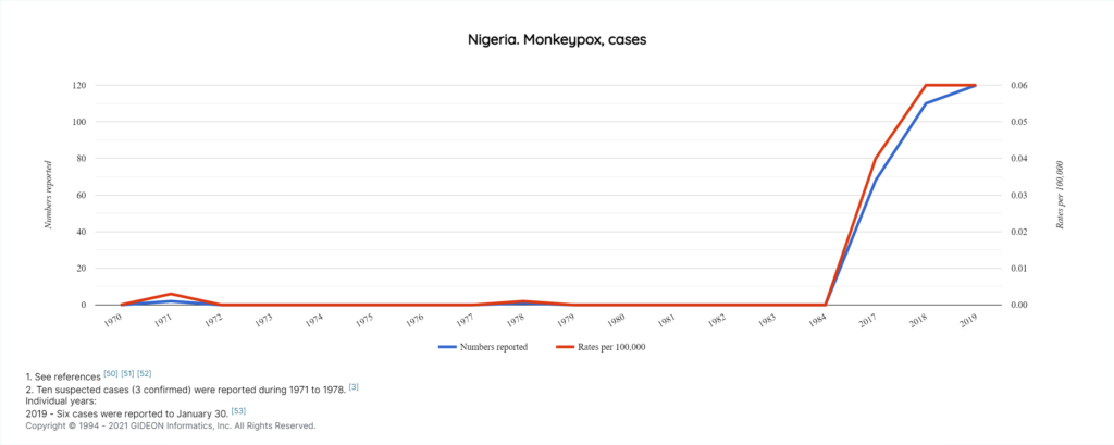
According to the GIDEON (Global Infectious Diseases and Epidemiology Network) country note on Monkeypox in Nigeria, there were six cross-border events of monkeypox from Nigeria to other countries and two notable outbreaks. In the latest one from 2017-2021, 446 people in Nigeria were infected with Monkeypox.


Monkeypox cases in Nigeria 1970 - 2019
Image: Graph illustrating Monkeypox cases in Nigeria 1970 – 2019. Copyright © GIDEON Informatics, Inc.


Diagnosing Monkeypox Accurately

Monkeypox has specific symptoms that distinguish it from other diseases. Swollen lymph nodes and a telltale rash help diagnose it more accurately, but misdiagnosis is possible. 

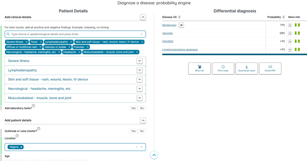
Let’s look at the illustration below. When we add patient travel history from Nigeria and the symptoms of monkeypox like fever, headache, generalized lymphadenopathy, skin lesions or rash, and others into the GIDEON infectious disease diagnostic probability engine, Varicella is also a strong contender. But the presence of ‘severe illness’ and the type of rash signifies a greater probability of Monkeypox.


GIDEON diagnostic probability engine. Differential diagnosis of Monkeypox
Image: GIDEON probability engine, differential diagnosis of Monkeypox. Copyright © GIDEON Informatics, Inc.


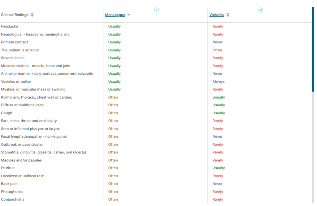
GIDEON’S side-by-side comparison of the clinical features of Monkeypox and Varicella helps narrow down and confirm the Monkeypox diagnosis based on patient presentation. 

GIDEON disease comparison table - Monkeypox vs. Varicella
Image: GIDEON disease comparison table – Monkeypox vs. Varicella. Copyright © GIDEON Informatics, Inc.


As Monkeypox has no cure, saving lives relies on early detection and control of spread.  Although there is some evidence that vaccination against smallpox may also prevent monkeypox, the Smallpox vaccine is no longer used. Another outbreak could happen anywhere, anytime.


Are we prepared to detect the next infectious disease that crosses our shores early? 

***

Detecting Foreign Infectious Diseases Early

Regarding the July 2021 Monkeypox case in Dallas, Dr. Philip Huang, the Director of the Dallas County Health and Human Services, stated, “This is another demonstration of the importance of maintaining a strong public health infrastructure, as we are only a plane ride away from any global infectious disease.”

Indeed, as the COVID-19 pandemic demonstrated, it is easy for emerging infectious diseases to spread rapidly worldwide. As seen in the Texas Monkeypox case, if patient travel history is considered at first presentation and symptoms are detected early, we can stop the spread of a disease-causing pathogen. 

Healthcare providers are our first line of defense against infectious diseases. Unfortunately, clinicians, nurses, emergency room workers, paramedics, and ambulance drivers in clinics and hospitals are also some of the first casualties from infection. The World Health Organization (WHO) recently reported that 115,000 healthcare workers have died from COVID-19. With the Delta variant on the loose, the pandemic is very much underway, and this number may rise. Protecting our borders and healthcare workers from emerging infectious diseases requires better access to advanced diagnostic tools with epidemiological data. 

When frontline clinicians have doubts about their initial assessment, they can conduct a differential diagnosis by comparing a patient’s symptoms with other diseases or consult infectious disease specialists. However, if they do not, or cannot, refer to a specialist or use an infectious disease platform for differential diagnosis (DDx), the disease-causing virus or bacteria may be misidentified and spread unchecked. 

***

The best way to catch emerging infectious diseases early is to equip frontline clinicians with comprehensive data on all reported infectious diseases, including:

  • a list of symptoms, 
  • the ability to compare conditions with similar presentations, and 
  • a history of global outbreaks and documented cases, 
  • country-specific notes on disease outbreaks. 


Did you like this article? Please share it on social media!

Check out more of our latest content here

Vaccine Heroes

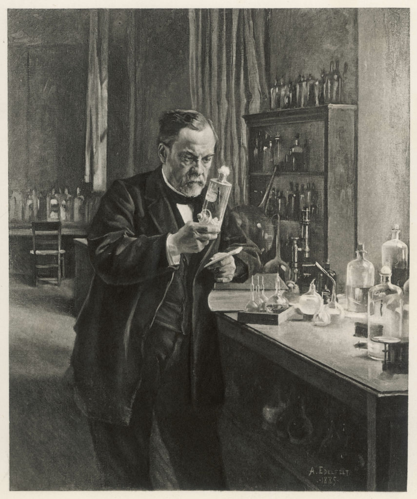
Louis Pasteur in his laboratory,1885
Louis Pasteur, the inventor of four vaccines, in his laboratory, 1885


The COVID-19 pandemic has been an eye-opener regarding the detrimental impact of microbial species on the human body. Vaccines act as vital tools for developing immunity against various infectious organisms through the recognition of targeted pathogens by the immune system. (Find more information on the mechanism of action of multiple types of vaccines here).

The initial development of vaccines resulted from the tireless efforts of many prestigious researchers who selflessly pursued the prevention of infectious diseases. Here is a brief sneak peek into the contributions of a few of these scientists whose invaluable efforts have saved millions of lives.


Portrait of vaccine hero Louis Pasteur

Louis Pasteur

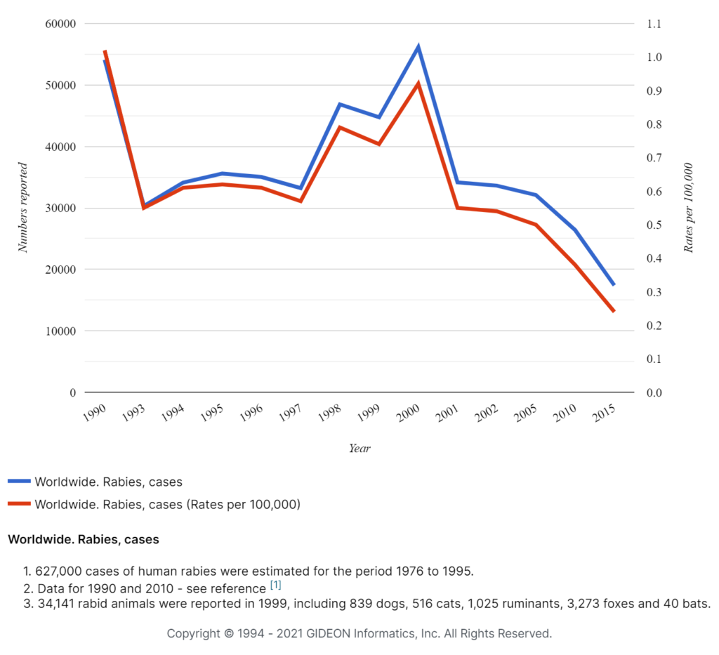
In the 1880s, Louis Pasteur developed vaccines for four potentially fatal infections, including Chicken Cholera, Anthrax, Swine Erysipelas, and Rabies. He was the first to introduce the use of live attenuated pathogens to develop immunity against the causative organisms (1). The vaccine for Chicken Cholera (Pasteurella multocida) was the first to be developed in a laboratory. Pasteur received several medals and honors, including the Leeuwenhoek Medal from the Royal Netherlands Academy of Arts and Sciences for his contributions to microbiology in 1895 (2).


Rabies cases and rates worldwide, 1990 – 2015

Rabies cases and rates worldwide, 1990 - 2015


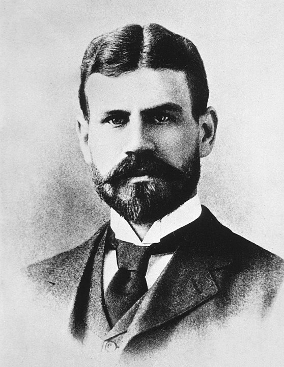
Waldemar Mordecai Wolffe Haffkine

Waldemar Mordecai Wolffe Haffkine

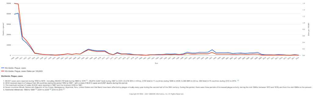
Waldemar Haffkine developed the first vaccines for Cholera and Plague, in the 1890s (3). Haffkine tested these inoculations on himself before initiating mass human trials. He conducted most of his studies in India, a hub of Cholera and Plague, and his monumental work saved the lives of millions of people.


Plague cases and rates 1948 – 2018

Plague cases and rates 1948 - 2018


Jesse William Lazear, 1866 - 1900

Jesse William Lazear

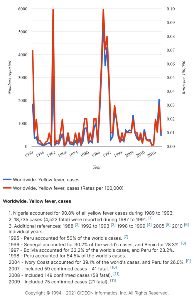
Dr. Jesse Lazear was an American physician who played a critical role in understanding the transmission of Yellow fever, a life-threatening viral infection (4). It was later revealed that he “allowed himself to be bitten by mosquitoes that had fed on the blood of patients with yellow fever,” which eventually led to his demise. His sacrifice was crucial in establishing the relationship between mosquitoes and Yellow fever, which later formed the basis of the development of key preventative strategies.


Max Theiler

Max Theiler

Max Theiler received the Nobel Prize in Medicine or Physiology “for his discoveries concerning Yellow fever and how to combat it” in 1951 (5). He pioneered the work on the development of a safe, standardized vaccine for the disease. In his studies, he used mice instead of rhesus monkeys, which were considered to be the main reservoir of the infection. Following this, mice continued to serve as standard tools for the study of zoonotic diseases by future researchers (6).


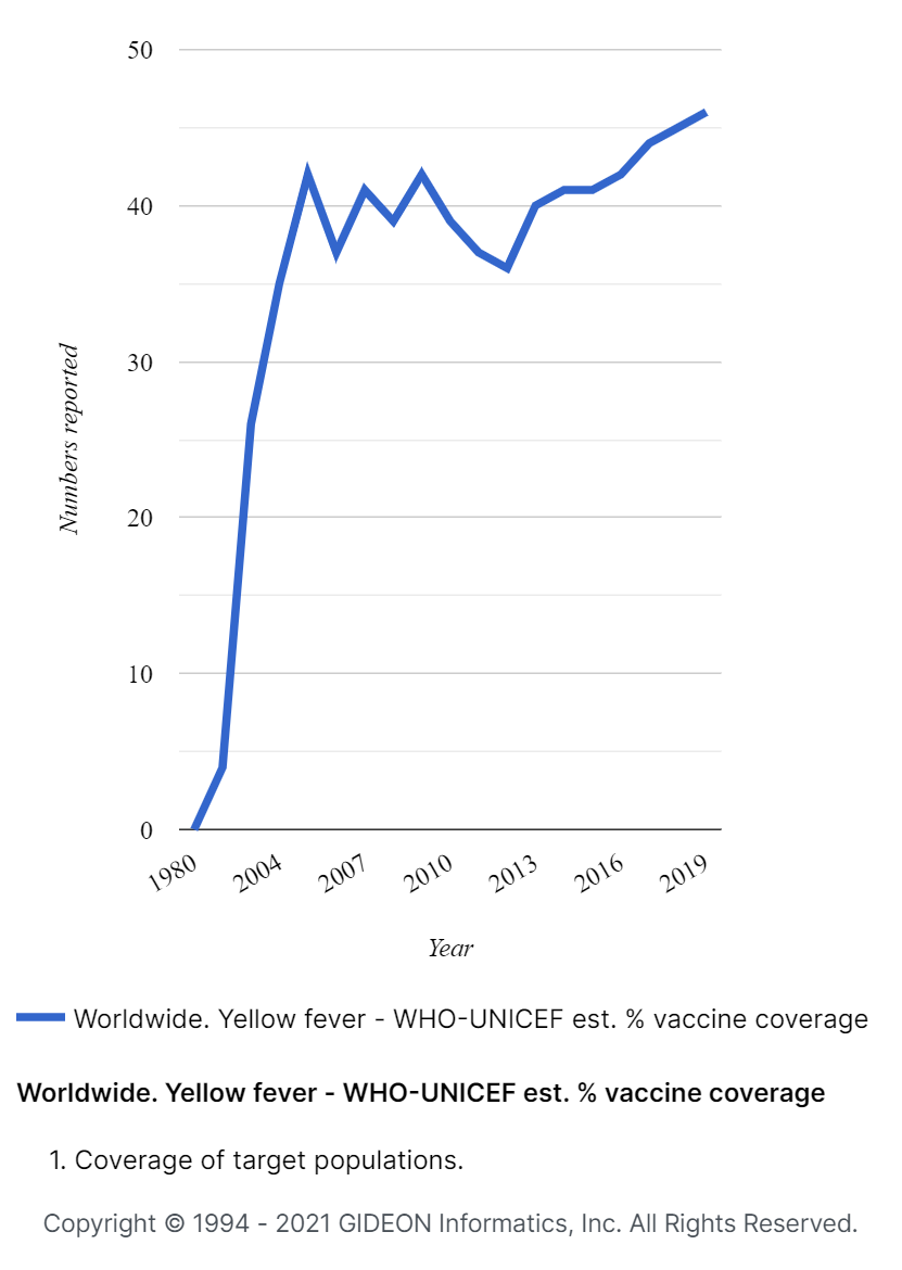
Yellow fever cases and rates worldwide, 1950 – 2016

Yellow fever cases and rates


Pearl Kendrick (left) and Grace Eldering. Photo credit: Michigan Women’s Hall of Fame

Grace Eldering & Pearl Kendrick

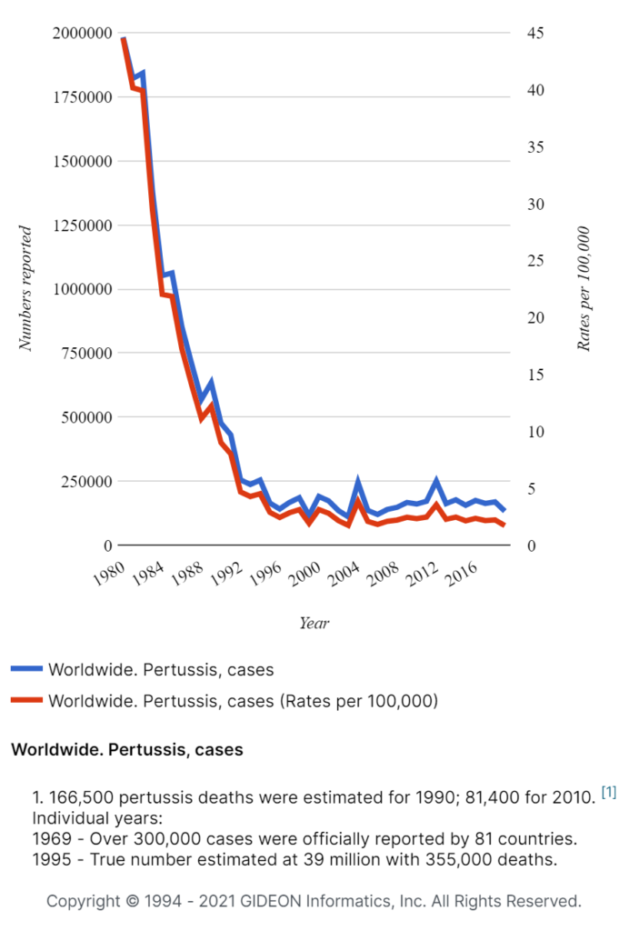
Both scientists conducted in-depth studies on Pertussis (whooping cough), which then became the basis of the development of a vaccine (7). Interestingly, both Grace Eldering and Pearl Kendrick suffered from whooping cough in their childhood, which was said to be the motivation behind their work. They were also involved in combining the Pertussis vaccine with those of Diphtheria and Tetanus to produce the DPT vaccine.


Pertussis cases and rates worldwide, 1980 – 2018

Pertussis worldwide 1980 - 2018


Portrait of John Franklin Enders

John Franklin Enders

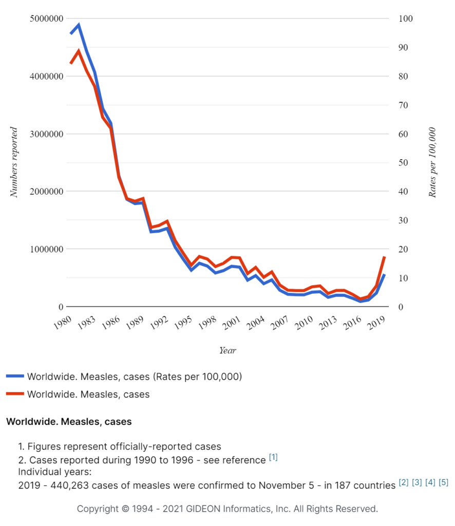
John Franklin Enders is referred to as “The Father of Modern Vaccines.” In 1954, he, along with Thomas H. Weller and Frederick C. Robbins, received the Nobel Prize in Physiology or Medicine for the successful in-vitro culture of the Poliomyelitis viruses (poliovirus) (8). Subsequently, Enders and his colleagues worked on developing a vaccine against the Measles virus, resulting in the availability of a live attenuated Measles virus vaccine and a deactivated Measles virus vaccine – marketed by Merck & Co. and Pfizer, respectively (9).


Measles cases and rates worldwide, 1980 – 2019

Measles worldwide cases and rates


The names mentioned above are just a few of the many scientists whose dedication, hard work, and intellect helped develop safe and effective vaccines, providing immeasurable contributions to our healthcare system.


Did you like this article? Share it on social media!

Check out more of our latest content here


References

  1. FLEMING A. Louis Pasteur. Br Med J. 1947 Apr 19;1(4502):517-22. doi: 10.1136/bmj.1.4502.517.
  2. “Leeuwenhoek Medal”, Royal Netherlands Academy of Arts and Sciences [Online]. Available. https://www.knaw.nl/en/awards/laureates/leeuwenhoekmedaille
  3.     Hawgood BJ. Waldemar Mordecai Haffkine, CIE (1860-1930): prophylactic vaccination against Cholera and bubonic Plague in British India. J Med Biogr. 2007 Feb;15(1):9-19. doi: 10.1258/j.jmb.2007.05-59.
  4.     Reed W, Carroll J, Agramonte A, Lazear JW. Classics in infectious diseases. The etiology of yellow fever: a preliminary note. Walter Reed, James Carroll, A. Agramonte, and Jesse W. Lazear, Surgeons, U.S. Army. The Philadelphia Medical Journal 1900. Rev Infect Dis. 1983 Nov-Dec;5(6):1103-11.
  5.     “Max Theiler Biographical”, The Nobel Prize [Online]. Available https://www.nobelprize.org/prizes/medicine/1951/theiler/biographical/
  6.     Norrby E. Yellow fever and Max Theiler: the only Nobel Prize for a virus vaccine. J Exp Med. 2007 Nov 26;204(12):2779-84. doi: 10.1084/jem.20072290.
  7.     Shapiro-Shapin CG. Pearl Kendrick, Grace Eldering, and the pertussis vaccine. Emerg Infect Dis. 2010 Aug;16(8):1273-8. doi: 10.3201/eid1608.100288.
  8.     “John F. Enders Biographical”, The Nobel Prize [Online]. Available https://www.nobelprize.org/prizes/medicine/1954/enders/biographical/
  9.     Katz SL. John F. Enders and measles virus vaccine–a reminiscence. Curr Top Microbiol Immunol. 2009; 329:3-11. doi: 10.1007/978-3-540-70523-9_1.

Tracking Dengue: An Interview With Alisa Aliaga-Samanez

“The rapid spread of Dengue could lead to a global pandemic, and so the geographical extent of this spread needs to be assessed and predicted. There are also reasons to suggest that transmission of Dengue from non-human primates in tropical forest cycles is being underestimated.”

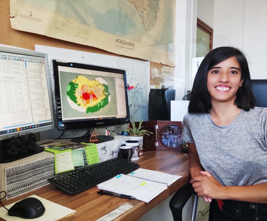
Alisa Aliaga-Samanez sitting by the computer
Alisa Aliaga-Samanez


Exactly one month ago, on June 7th, PLOS Neglected Tropical Diseases published a research article Worldwide dynamic biogeography of zoonotic and anthroponotic Dengue. The study is the first high-resolution analysis of how the risk of Dengue transmission has been changing geographically since the late 20th century, indicating the virus (DENV) has been making a home in previously low-risk areas, potentially due to global warming and deforestation. 

We spoke with the corresponding author Alisa Aliaga-Samanez, who worked alongside Marina Cobos-Mayo, Raimundo Real, Marina Segura, David Romero, Julia E. Fa, and Jesús Olivero, to learn more about the importance of this study and her experience working with GIDEON data. 


How did you find out about GIDEON?  

We got to know GIDEON thanks to an article published in PNAS by Kris A. Murray and colleagues entitled “Global biogeography of human infectious diseases.”


What were the reasons behind choosing the GIDEON database as one of your data sources?

We know that GIDEON is one of the most complete data sources worldwide on zoonoses, so we wanted to use it to build our database. For that reason, the project where I work at the University of Malaga funded our access to GIDEON.


What is the importance of biogeography studies like this one to public health management?  

Biogeographical studies, through modeling, applied to pathogens, allow us to understand the distribution of infectious diseases. The occurrence of disease cases is related to social factors but also to environmental variables that determine the degree to which certain environments favor the occurrence of disease, even where it has not been recorded. Thanks to the tool we use in our study, we are able to propose different management strategies depending on which factors favor the risk of transmission in different regions of the world. We took into account three possible biogeographical scenarios related to Dengue transmission risk: (1) zones with favorable conditions for viruses and vectors, (2) favorable conditions for virus only, and (3) favorable conditions for vectors only. Besides, our biogeographical approach helped us to analyze the extent of the areas where non-human primates could be involved in sylvatic Dengue cycles.


Do you believe the risks of the Dengue pandemic are exacerbated by global warming?  

We think global warming may be one of the factors that could be favoring vectors to adapt to new environments. A study published by Messina and colleagues in 2019 concluded that the World’s population at risk of Dengue could experience an almost 60% increase by 2080. In addition, they suggested that outbreaks could reach areas in continents such as Australia, Argentina, Japan, eastern China, and southern Europe.

Melting ice
Photo by William Bossen on Unsplash


In your experience, how effective study such as yours at influencing change in government and health policies, and do you think work like this will be taken more seriously following the COVID-19 pandemic?  

We have seen that governmental bodies such as WHO or CDC rely on scientific studies to manage risk areas, but we feel that this work is being limited in Africa, for example. On the other hand, some diseases already have a vaccine available, such as yellow fever, but its application might not be well managed in some African countries, where large outbreaks occur. In the case of Dengue, a vaccine has already been developed, but it is only licensed for people aged 9-45 years in some countries. The WHO recommends that the vaccine should only be administered to people with confirmed previous Dengue virus infection. As we show in our paper, Dengue is currently spreading together with its vectors. The COVID-19 pandemic should serve to demonstrate that global pandemics are possible and, so, must be prevented.


What value did having access to global spatio-temporal data add to your study?  

GIDEON helped us to get quick access to the available information on the disease. This allowed us to build our databases in a short time and to validate our models with recent cases in order to assess the reliability of the tool we use and to predict new areas favorable for new Dengue cases.


What are your thoughts regarding data availability on sylvatic transmitted Dengue?  

Most of the available data on human cases do not differentiate whether they were caused by sylvatic or urban transmission. Some research studies in certain local areas may be able to determine this, but globally there is no such information. Our biogeographic outputs related to Africa and Asia are consistent with the scarce information available and provide the context in which on-the-ground prospections on sylvatic Dengue should be addressed. This is especially important in South America, where sylvatic Dengue has not been detected yet (although the presence of sylvatic Yellow Fever and other evidences are starting to suggest its existence).

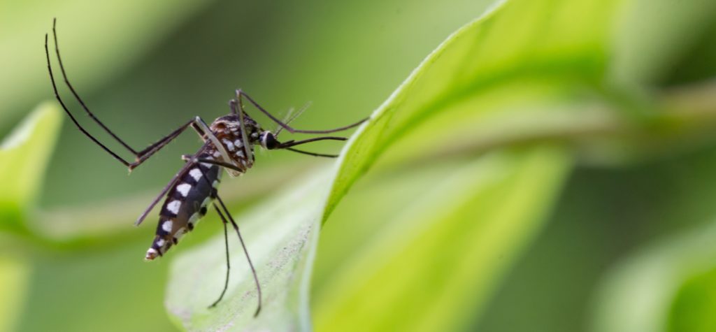
Aedes aegypti Mosquito. Close up a Mosquito Mosquito on leaf,Mosquito Vector-borne diseases,Chikungunya.Dengue fever.Rift Valley fever.Yellow fever.Zika virus.
Aedes aegypti mosquito, the vector of Dengue


What areas do you think GIDEON could improve to make our data more useful to you in the future?  

We appreciate the great work GIDEON does in collecting data globally. We found the new GIDEON interface very useful. Perhaps it would make it easier in the future, if it is possible, to distinguish between imported and autochthonous cases within the database. It is true that in many cases the database does mention it but not for all countries.


About Alisa:

Alisa Aliaga-Samanez has a degree in Biology from the Federico Villarreal University (Peru) and a Master’s degree in Biodiversity and Environment from the University of Malaga (Spain). Currently, she is working at the Animal Biology Department of the University of Malaga, being part of the Biogeography, Diversity and Conservation Group,  developing her Ph.D. thesis. The thesis is focused on the study of primate biogeography, applied to conservation and human health. She is currently mapping vector-borne zoonotic diseases through global distribution modeling. 

Aliaga uses high-resolution global maps and the most up-to-date databases to analyze geographical changes in the risk of zoonotic disease transmission. In addition, in these analyses, she considers the biogeographical contribution of primates in increasing the risk of transmission. She seeks to determine the potential natural range of endemic and emerging zoonotic diseases in the world, with the aim of suggesting specific management strategies according to the spatial distribution of risk factors.

Click here to read the open-access article: Worldwide dynamic biogeography of zoonotic and anthroponotic Dengue 


Did you like this article? Share it on social media!

Check out more of our latest content here

Pathogen of the month: Mycobacterium tuberculosis

Mycobacterium tuberculosis (M. tuberculosis), a non-motile, obligately aerobic, intracellular bacterium known to cause Tuberculosis (TB), was discovered by Robert Koch in 1882 [1]. TB primarily affects the lungs along with the abdomen, bones, nervous system, reproductive system, liver, and lymph glands [2].

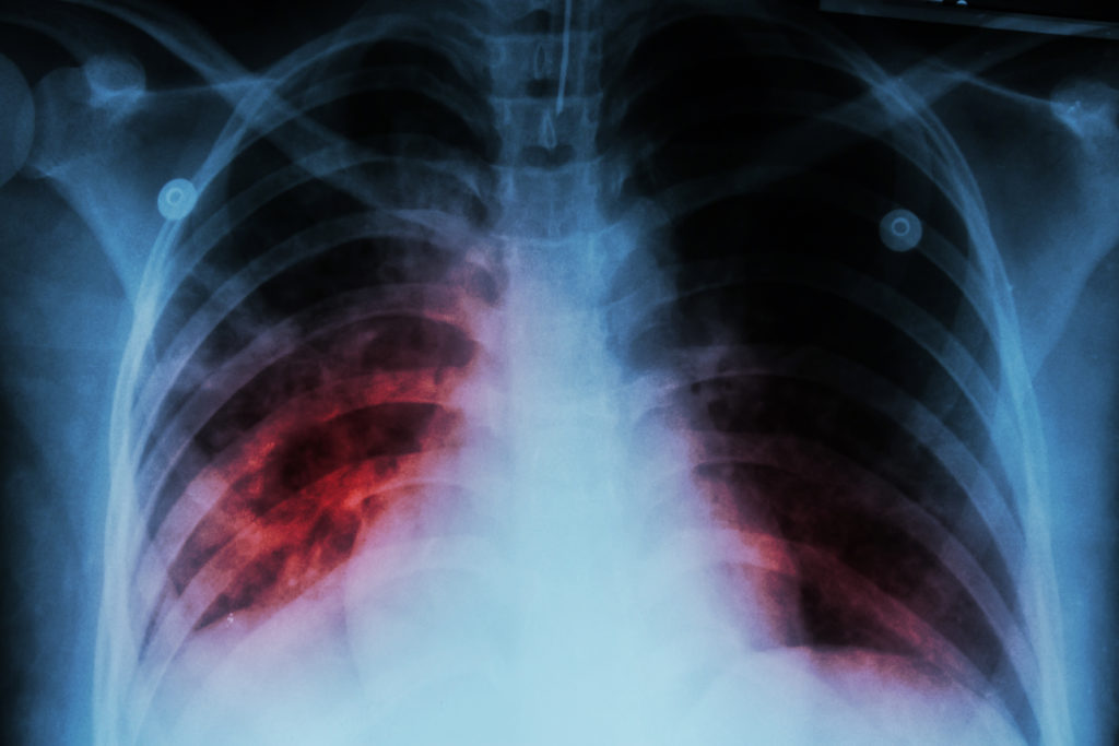
Pulmonary Tuberculosis ( TB ) : Chest x-ray show alveolar infiltration at both lung due to mycobacterium tuberculosis infectio
Pulmonary Tuberculosis ( TB ): Chest x-ray shows alveolar infiltration at both lungs due to Mycobacterium tuberculosis infection


Global Burden of Tuberculosis

The World Health Organization (WHO) has reported a 22% decrease in TB-related mortality between 2000-2015, with a gradual 1.5% decrease in the annual rate. However, despite this trend and successful control of disease transmission, TB continues to have a significantly higher rate of morbidity. In 2015, six countries (China, India, Nigeria, Pakistan, Indonesia, and South Africa) accounted for 60% of TB-related deaths [3].


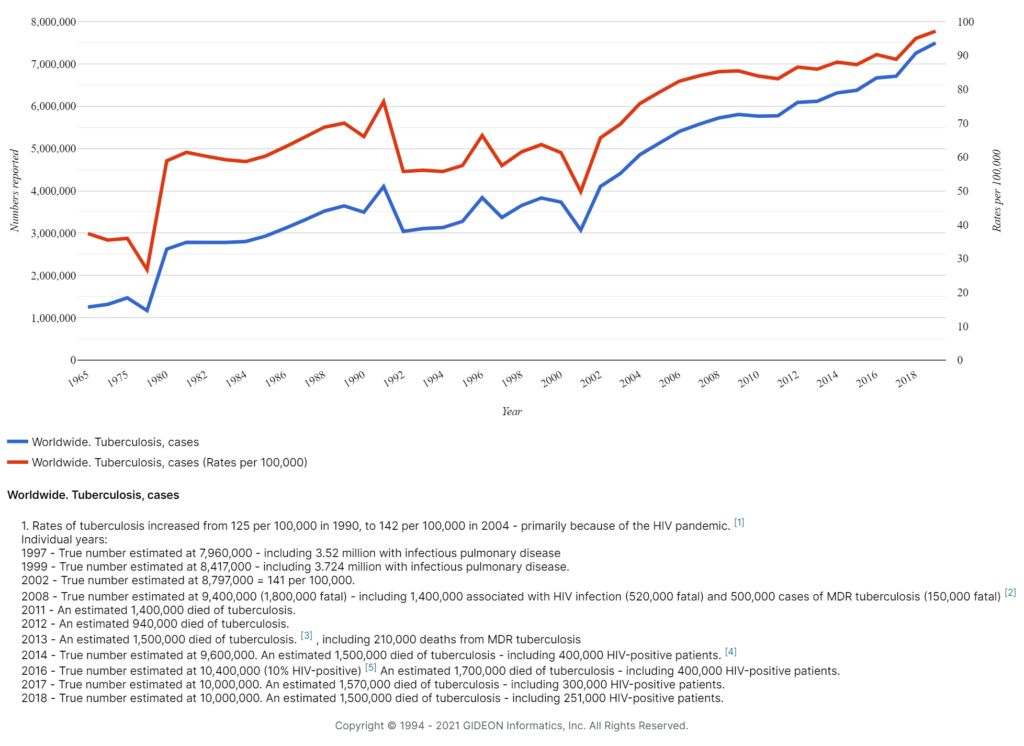
Global cases of Tuberculosis between 1965-2019

Worldwide Tuberculosis cases and rates, 1965 - today


Pathogenicity of Mycobacterium tuberculosis

Mycobacterium tuberculosis is transmitted in the form of droplet nuclei exhaled by individuals affected with laryngeal/pulmonary TB. It enters the body via the nasal cavity/mouth and travels to the alveoli of the lungs, where it recruits macrophages to the lung surface, in turn transporting the bacteria to the deeper tissues [4]. Another round of macrophage recruitment to the originally infected locus forms an organized aggregate of differentiated macrophages and immune cells called a granuloma. The infected granuloma undergoes necrosis, promoting bacterial growth and transmission to the next host [5].

Diagnosis of TB

Some patients with TB may present with non-specific findings such as anemia, weight loss, fever of unknown origin, and fatigue; while others may be asymptomatic and show no abnormalities on physical examination [6]. Thus, the physician should collect body fluids/tissues for Acid-Fast bacilli (AFB) smear and culture.  Only a positive culture can confirm the diagnosis of TB.

Confirmatory and diagnostic tests for TB:

– Culture, followed by Ziehl-Neelsen (AFB) staining

– A Chest X-ray to confirm the diagnosis in case of positive culture, indicating active disease

– Gene-based tests and nuclear amplification to identify the bacterial strains using DNA-based molecular techniques, such as GeneXpert [7].


Ziehl-Neelsen staining

AFB staining is the traditional method of TB diagnosis, as it is inexpensive and provides rapid results [8]. Mycobacterium species retain dyes when heated and treated with acidified organic compounds. The most common acid-fast staining method for M. tuberculosis is the Ziehl-Neelsen stain method, in which a specimen is fixed, stained with carbol-fuchsin dye, and decolorized with an acid-alcohol mixture. After counter-staining the smear with methylene blue or a similar dye, AFB appear red against a contrasting blue background [9].  In general, a sputum sample must contain at least 10,000 organisms/mL to visualize these bacteria at 100x magnification. 

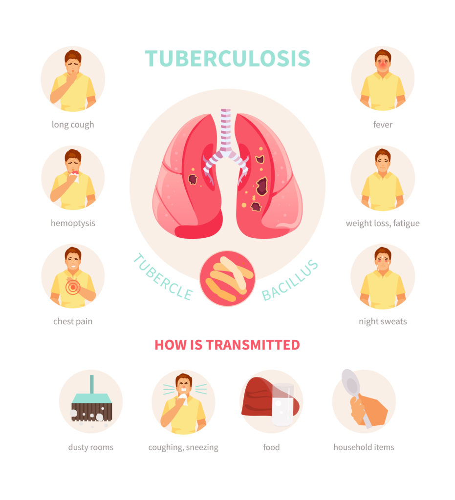
Symptoms and ways of infection of tuberculosis. Medical vector infographics, poster

Latent TB vs. Active TB

Latent TB represents the condition where the body’s immune system restricts the growth of M. tuberculosis bacterium, making the individual appear asymptomatic [10].

An individual with latent TB infection shows

– no symptoms.

– is not infectious (cannot spread TB).

– tests positive for TB blood/skin tests.

– may eventually develop active TB if the immune system weakens.

Active TB represents a condition where the body’s immune system is unable to restrict the growth of M. tuberculosis, rendering patients both ill and contagious. 

The symptoms of TB depend on the affected area.

a. General symptoms include:

  1. Night sweats
  2. Weight loss
  3. Prolonged fever
  4. Loss of appetite
  5. Fatigue

b. Symptoms of Pulmonary TB (infected lungs):

  1. Shortness of breath, which progressively worsens
  2. A persistent cough that produces phlegm and sometimes blood, persisting  > 3 weeks. 

c. Symptoms in the case when other areas of the body are infected:

  1. Swellings in the neck or other regions
  2. Pain in a joint or affected bone
  3. Abdominal pain
  4. Headache
  5. Confusion
  6. Seizures


Treatment

According to the World Health Organization (WHO), first-line treatment for TB may include combinations of five essential drugs: Rifampicin (R), Isoniazid (H), Pyrazinamide (Z), Ethambutol (E), and Streptomycin (S) [11].  Patients with TB undergo a standardized treatment for 6 to 9 months, including an initial two-month course of Rifampicin, Isoniazid, Pyrazinamide, and Ethambutol, followed by another 4-month course of Isoniazid and Rifampicin [12].

For patients with Multi Drug-Resistant Tuberculosis (MDR-TB), directly-observed therapy (DOT) is used. In DOT, drugs are administered at least six days/week under the direct observation of the physician [13, 14]

BCG vaccine

Bacillus Calmette-Guerin (BCG), a vaccine for TB, was introduced in 1921 to control tuberculosis in humans. It is administered at birth, primarily in regions with a high disease burden, such as India, South Africa, and Pakistan [15]. Widespread immunization using BCG vaccine has facilitated a reduction in TB cases globally [16].


Risk factors

Five-to-ten percent of people with latent TB who do not receive appropriate treatment will eventually develop active TB disease [17]

Individuals at a higher risk of contracting TB include:

– Those who have traveled to or are living in a country with a high prevalence of TB. 

– Those living in crowded conditions

– Those who have been in close contact with a person infected with TB

– Children ages <= 5 years who have tested positive for TB

– People who reside or work with persons at high risk for TB, such as those in hospitals, correctional facilities, homeless shelters, residential homes for HIV-infected individuals, and nursing homes

Additionally, immune dysfunction associated with diabetes mellitus, HIV infection, cancer chemotherapy, malnutrition, and advanced age is associated with an increased risk of contracting TB. 

Additional conditions associated with high risk for tuberculosis include silicosis, substance abuse, malignancy, organ transplantation, corticosteroid therapy, Crohn’s disease, and rheumatoid arthritis.

Tuberculosis and HIV

Co-infection by TB and HIV places a diagnostic and therapeutic burden on the health care system. HIV infection has been shown to increase the risk of reactivation of latent TB by 20-fold. [18].


Did you like this article? Share it on social media!

Check out more of our latest content here


References

  1.     K. R, Die Aetiologie der Tuberculose [The aetiology of Tuberculosis.], Berlin: Berliner Klinische Wochenschrift, 1882.
  2. F. K. Dye C, Disease Control Priorities in Developing Countries, New York:: Oxford University Press, 2006.
  3. Z. J. B. Q. H. H. B. L. Y. J. L. Q. L. J. Pan Z, “The Gap Between Global Tuberculosis Incidence and the First Milestone of the WHO End Tuberculosis Strategy: An Analysis Based on the Global Burden of Disease 2017 Database.,” 2020.
  4. CDC, How TB Spreads, CDC, 2016.
  5. G. M. J. Jr., “Microbial pathogenesis of Mycobacterium Tuberculosis: dawn of a discipline,” Cell, no. 104, pp. 477-485, 2001.
  6. J. D. E, Mycobacterial diseases of the lung and bronchial tree: Clinical and laboratory aspects of Tuberculosis, Boston: Brown and Company, 1974.
  7. [Online]. Available: https://www.ncbi.nlm.nih.gov/books/NBK441916/.
  8. B. J. R. Elizabeth A. Talbot, Molecular Medical Microbiology, 2015.
  9. R. l. Kradin, Diagnostic Pathology of Infectious Disease, 2018.
  10. World Health Organization, Guidelines on the management of latent Tuberculosis infection, Geneva: WHO, 2015.
  11. World Health Organization, Implementing the WHO Stop TB Strategy: A Handbook for National Tuberculosis Control Programmes.
  12. Gideononline, “www.gideonoline.com,” Gideon, 2021. [Online]. Available: https://app.gideononline.com/explore/diseases/Tuberculosis-12470.
  13. A. F. Z. L. F. E. Terracciano E, [Tuberculosis: an ever present disease but difficult to prevent], Ig Sanita, 2020.
  14. M.-R. K. R. R. C. R. Chaulk CP, Eleven years of community-based directly observed therapy for Tuberculosis, JAMA, 1995.
  15. “www.journals.plos.org,” [Online]. Available: https://journals.plos.org/plosmedicine/article/figures?id=10.1371/journal.pmed.1001012.
  16. Z. a. Lancione, “Using data science to improve knowledge around a century old vaccine,” The BCG Atlas, 2020.
  17. CDC. [Online]. Available: https://www.cdc.gov/tb/topic/basics/risk.htm.
  18. G. C. G. R. N. P. Getahun H, “HIV infection-associated Tuberculosis: the epidemiology and the response,” Pubmed, 2010.

Diagnosing Emerging Infectious Diseases Early: How Epidemiology Can Help Clinicians on the Frontline

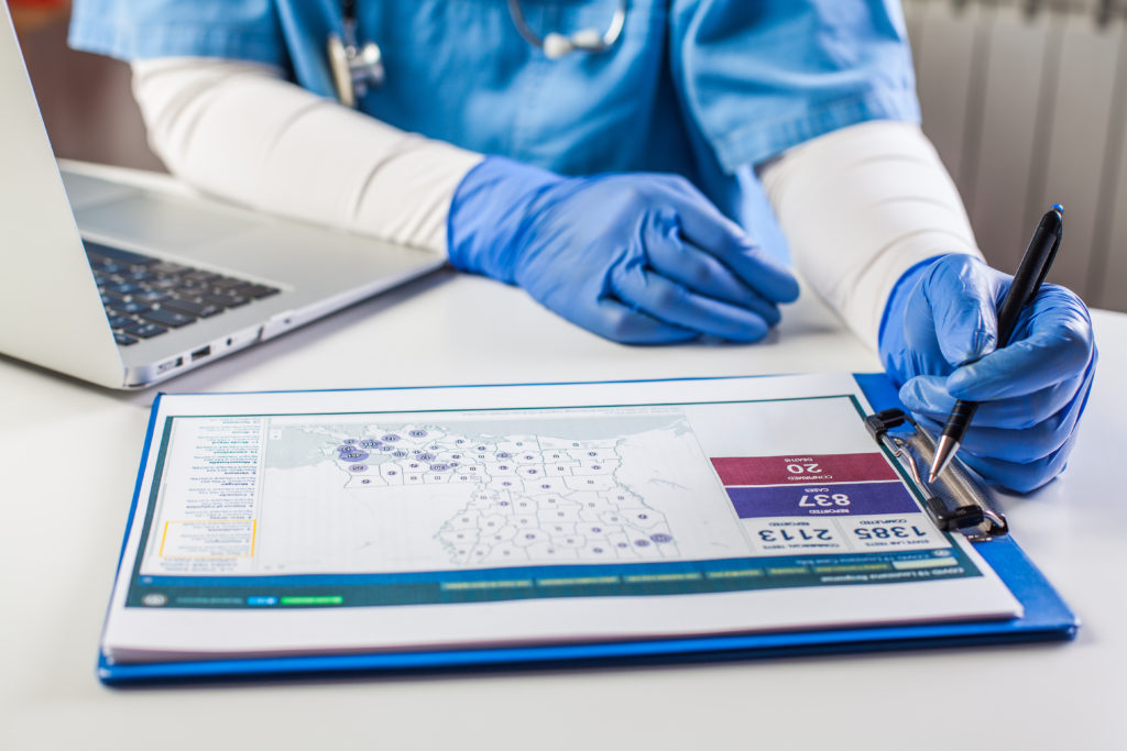
Doctor wearing protective gloves working on laptop computer,analyzing Coronavirus info data,COVID-19 response case info,U.S. state rank cases per capita,percent infected info and population numbers
Patient travel history or epidemiological data is the missing link in the differential diagnosis


by Chandana Balasubramanian

In 2019, a crew of nine pilots and astronauts broke a world record. They flew around the Earth in just 46 hours. More incredible is that they did not fly in a never-before-seen, advanced aircraft prototype. They flew a commercially available jet plane.

The future is already here.

It’s a small world; it’s getting smaller by the minute

Based on the new world record, it could take less than a weekend for an emerging infectious disease to spread all over the globe. And chances are, it may take a week or more before it gets detected based on the incubation period.

Unfortunately, healthcare providers at the frontlines of infectious disease management face a significantly higher risk of infection. The risk extends beyond healthcare workers to their families and communities.

As the world grapples with the impact of COVID-19 and its mutations, it’s a good time to ask: What can health systems worldwide do to detect emerging infectious diseases imported from other countries early?

1. Think beyond the travel ban

Recent research empirically demonstrated that local outbreaks of various Infectious Diseases could “quickly spread to other countries through the international movement of people and goods, with potentially disastrous health consequences [1].”

While this fact may not be news to clinicians and Infectious Disease specialists, the study also shows a close spatial dependence between the health conditions in one country and another – a spillover effect. The study used GIDEON (Global Infectious Disease and Epidemiology Online Network), a database covering all Infectious Disease outbreaks.

An epidemic in one country can become a pandemic in others – irrespective of travel and other physical barriers to entry. Studies of previous epidemics show that even a 90% travel restriction between countries merely delays the arrival of an emerging infection by a few weeks. Another study by Quilty et al. reported that airport-based screening measures to detect COVID-19 missed 46% of cases because of the incubation period [2].

So, while a travel ban and thermal screening can help a country buy some time to prepare for an outbreak, epidemic, or pandemic, they cannot stop or prevent a new infection from spreading to foreign shores.

***

2. Record travel history as standard protocol

Travel has always been one of the fastest ways to introduce a pathogen to a new environment. And as two clinicians, Trish Perl and Connie Savor Price, argue in a recent ‘Annals of Internal Medicine’ article, travel history must be treated as the fifth vital sign in emergency rooms and all physician evaluations [3].

The doctors make a strong case that including a patient’s travel history as part of a vital signs check can “help put symptoms of infection in context and trigger us to take a more detailed history, do appropriate testing, and rapidly implement protective measures.”

Monkeypox in the UK, 2021

For example, in May 2021, the World Health Organization received notification from the United Kingdom of a confirmed case of monkeypox in an individual who had just traveled from Nigeria. Monkeypox has an incubation period of six to thirteen days, but according to WHO, it can range anywhere from five to twenty-one days. Eventually, the infection spread to another family member, and they were isolated. Differential diagnosis considerations for monkeypox include chickenpox, measles, bacterial infections, scabies, syphilis, and medication-associated allergies. In such a case, taking the patient’s travel history can help healthcare workers take the necessary precautions even before the PCR results.

COVID-19 in the United States, 2019

The first case of COVID-19 in the US was reported in Washington when the patient returned from Wuhan, China. Based on the patient’s travel history and symptoms, healthcare professionals could isolate and send clinical specimens to be tested by the CDC overnight. Hospitals in the United States were already on alert for patients from Wuhan presenting with symptoms, and testing could be prioritized accordingly.

Ebola in the United States, 2014

Let’s look at an example where a patient’s travel history would have helped protect healthcare professionals. In 2014, a man traveled from West Africa and admitted himself into a hospital in Dallas with fever, abdominal pain, dizziness, headache, and nausea. Without an integral piece of the puzzle – his travel history – he was treated for sinusitis and sent home. The hospital suspected Ebola only when he returned three days later with persistent fever, abdominal pain, and diarrhea. Unfortunately, within this time, this patient had infected healthcare professionals, ambulance transport personnel, and the patient’s caregivers.

Monkeypox and Ebola are not as contagious as COVID-19 and its variants, and Ebola is not contagious until symptoms appear, making containment easier. But emerging infectious diseases and their variants might be.

Infectious Disease specialists, clinicians, researchers, and medical librarians will need to be vigilant against the next outbreak. Epidemiological data plays an integral role in facilitating improved clinical decisions and saved lives.

***

3. Identify initial cases of known diseases in new settings

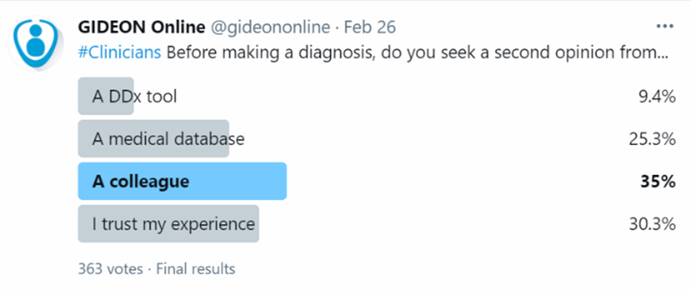
In a GIDEON survey of 363 clinicians in the US, UK, and Canada, 35% stated that they would consult a colleague for a second opinion before making clinical decisions. As a close second, 30% indicated that they trust their judgment. This means that 65% of the survey respondents trusted human judgment over Point-of-Care tools.

Twitter survey of clinicians and use of differential diagnosis tools

But the stakes are higher when dealing with highly transmissible emerging infections. The importance of first-time diagnosis accuracy is compounded due to the rising urgency to prevent the next epidemic or pandemic.

Consider the dramatic difference in transmission rates between SARS-CoV-2 and its variants:

  • The B.1.1.7, the ‘Alpha’ SARS-CoV-2 variant, is 43% to 90% more transmissible than its predecessor and led to a surge in hospitalizations across the UK and 114 more countries in a mere few months [4].
  • 1.617.2 or the ‘Delta’ variant is estimated to be 40% to 60% more infectious than the Alpha, estimated by disease modelers at Imperial College, London, with an R0 as high as 8 [5].

Here are some comparisons of how newer, emerging pathogens and their variants compare to older, Infectious Diseases.

Pathogen Transmissibility Rate (R0)
B.1.617.2, SARS-CoV-2 Delta variant 5-8
B .1. 1. 7, SARS-CoV-2 Alpha Variant 4-5
SARS-CoV-2 (COVID-19) 2.5
SARS-CoV 2.4
Measles 1.5 (1.5-2.0)

In other words, an outbreak may already be well underway before an Infectious Disease specialist is consulted for assistance on differential diagnosis or a medical librarian is requested for location-specific disease symptoms.

As pathogens mutate, traditional methods of differential diagnosis need an upgrade. Clinicians, Infectious Disease specialists, and researchers need data from local outbreaks anywhere in the world at their fingertips to help drive decision-making and advance the global effort against Infectious Disease.

***

4. Use a differential diagnosis (DDx) tool like GIDEON’s First Case Scenario to identify Infectious Diseases – faster and more accurately

Drs Perl and Price champion the need for greater access to digital resources that integrate electronic health records with patient travel histories and can “suggest specific diagnoses in febrile returning travelers.”

One of the more well-known DDx tools is GIDEON with its First Case Scenario feature, created in partnership with the World Health Organization (WHO) after the West Nile Fever outbreak in the United States.

Using a DDx platform such as GIDEON helps:

  • narrow down possibilities,
  • lead to a faster result,
  • reduce the margin of error at the point-of-care, and
  • elevates peer-to-peer knowledge sharing on a global scale

Why is this important? Because, for example, in respiratory viral illnesses, early detection is the critical step to mitigate disease transmission but is often delayed [3]. Depending on the type of pathogen, this could lead to a greater number of hospitalizations, more morbidity, a burden on healthcare systems, and have significant ramifications on a country, its people, and the economy.

Locations marked with pins on world map, global communication network, closeup. Asking patients about their travel history can help prevent emerging infectious diseases introduction into country
Asking patients about their travel history can help prevent emerging infectious diseases introduction into the country

Having a differential diagnosis platform that incorporates a patient’s travel history can make a huge difference in how the world manages emerging infectious diseases.

Here’s an example. Suppose a patient presents with elevated body temperature, severe headache, chills, myalgia, diarrhea, and malaise.

These are nonspecific presentations and could be representative of a variety of diseases.  With international transmission now the norm, no clinician can be expected to keep track of every single emerging disease and its symptoms.

Example: Diagnosing Ebola using a DDx platform

Step 1: Focusing on most likely diseases based on symptoms and travel information
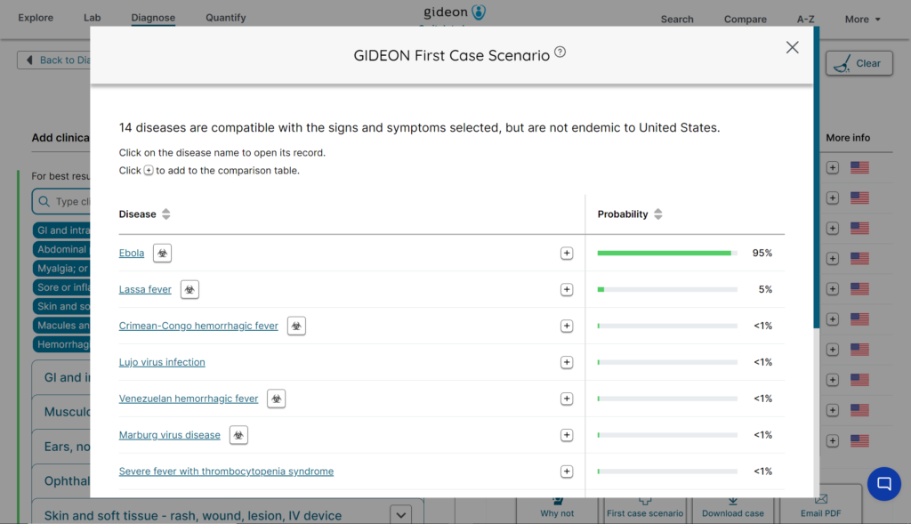
Entering a patient’s symptoms and the locations and dates of travel in a tool like GIDEON’s Bayesian analysis-driven Probability engine can help identify what diseases are most likely to correspond to the data entered. The illustration below shows Ebola as a high probability based on the patient’s symptoms and travel location.

Step 2: Conduct a differential diagnosis
The screenshot of the First Case Scenario feature below shows a 95% probability that the patient has Ebola. What if there were fewer symptoms at presentation, the likelihood of Ebola was 65%, and another disease was 25% probable? You could conduct a differential analysis by comparing the two disease symptoms on the platform, download the comparison, and order the requisite laboratory tests to confirm.

Step 3: First Case Scenario

Imagine it is 2014, and you haven’t heard of Ebola. A patient walks in with the symptoms listed above. You enter the symptoms and the patient’s travel history. Using GIDEON’s First Case Scenario, you can determine how likely it is that your patient is the first in the country to present with Ebola.

GIDEON web application screenshot displaying 95% probability of Ebola in the First Case Scenario feature
Illustration of diagnosing Ebola in a Point-of-Care setting (Screenshot: GIDEON First Case Scenario DDx tool)

***

5. Train an army of global clinicians to battle Infectious Diseases

Based on a GIDEON survey of 230 clinicians in the US, UK, and Canada, while clinicians were open to using a DDx tool to help diagnose Infectious Diseases, a lack of budget was the primary reason they did not.

One physician even stated, “I would use them every day if my institution would offer.”

But an interactive platform with a robust database of Infectious Disease symptoms that incorporates patient locations, exposure to disease-causing elements, and comparisons between two or more similar diseases can offer benefits beyond what a seasoned clinician can accomplish.

It can train the next generation of Infectious Disease-fighting doctors and healthcare professionals. For example, take GIDEON’s step-by-step Bayesian analysis toolkit. Teaching institutions, medical librarians, medical students, residents, researchers, and more can use DDx tools to help hone their diagnoses of emerging as well as well-known infectious diseases.

The tool helps you list symptoms, patient travel information (if any), and any exposure to disease-causing elements (if known). For example, the patient ate chicken in a region that recently had a Salmonella outbreak.

The tool offers a list of probable diseases in descending order of probability. It helps that the tool is dynamic because what if the patient forgot a symptom and told you about it later? A new list of probable diseases is re-calculated automatically. An added benefit is that the DDx tool is integrated with the First Case Scenario to determine if a patient’s symptoms are the first in a specific location.

Health systems and medical colleges and universities may benefit greatly from such a diagnostic solution.

Conclusion

War often provides an opportunity for innovation. After all, the internet was invented because computers at the time were enormous, and it was incredibly difficult to physically transport military intel from the United States to soldiers deployed around the world [6]. And clinicians are actively in a battle against the spread of infectious pathogens.

A global platform that offers timely location-specific intelligence about emerging infectious diseases and helps speed up clinical decisions is invaluable to future-proof the world against outbreaks, epidemics, and pandemics and save thousands of lives.

Did you like this article? Share it on social media!

Check out more of our latest content here

References

[1] R. Desbordes, “Spatial dynamics of major Infectious Diseases outbreaks: A global empirical assessment,” J. Math. Econ., vol. 93, no. 102493, p. 102493, 2021.

[2] B. J. Quilty, S. Clifford, S. Flasche, R. M. Eggo, and CMMID nCoV working group, “Effectiveness of airport screening at detecting travellers infected with novel coronavirus (2019-nCoV),” Euro Surveill., vol. 25, no. 5, 2020.

[3] T. M. Perl and C. S. Price, “Managing emerging Infectious Diseases: Should travel be the fifth vital sign?” Ann. Intern. Med., vol. 172, no. 8, pp. 560–561, 2020.

[4] N. G. Davies et al., “Estimated transmissibility and impact of SARS-CoV-2 lineage B.1.1.7 in England,” Science, vol. 372, no. 6538, p. eabg3055, 2021.

[5] Scientific Advisory Group for Emergencies, “Imperial College London: Evaluating the roadmap out of lockdown – modelling Step 4 of the roadmap in the context of B.1.617.2 (Delta), 9 June 2021,” Gov.uk, 14-Jun-2021. [Online]. Available: https://www.gov.uk/government/publications/imperial-college-london-evaluating-the-roadmap-out-of-lockdown-modelling-step-4-of-the-roadmap-in-the-context-of-b16172-delta-9-june-2021. [Accessed: 15-Jun-2021].

[6] B. Tarnoff, “How the internet was invented,”  The Guardian, 15-Jul-2016.

Preventing Foodborne Illness

Food quality control expert inspecting specimens of groceries in the laboratory
Food quality control expert inspecting groceries in the laboratory


Since 2019, World Food Safety Day is celebrated each year on the 7th of June,  to promote awareness regarding the need to develop and maintain food safety standards. These efforts are being done both to reduce the burden of foodborne disease and to minimize its impact on socio-economic systems.

Foodborne diseases constitute a major health issue worldwide. The World Health Organization (WHO) estimates that approximately 10% of the global population is infected by foodborne pathogens each year (1). Similar to other diseases, the severity of the foodborne illness varies with the type and the amount of exposure to the causative microorganism, as well as the disease-fighting potential of the affected individual.


Clinical Presentation of Foodborne Illnesses

Foodborne illnesses might manifest as acute diarrhea, nausea, etc., which usually resolves within 7 to 10 days; or as chronic and potentially fatal diseases such as botulism or typhoid fever.

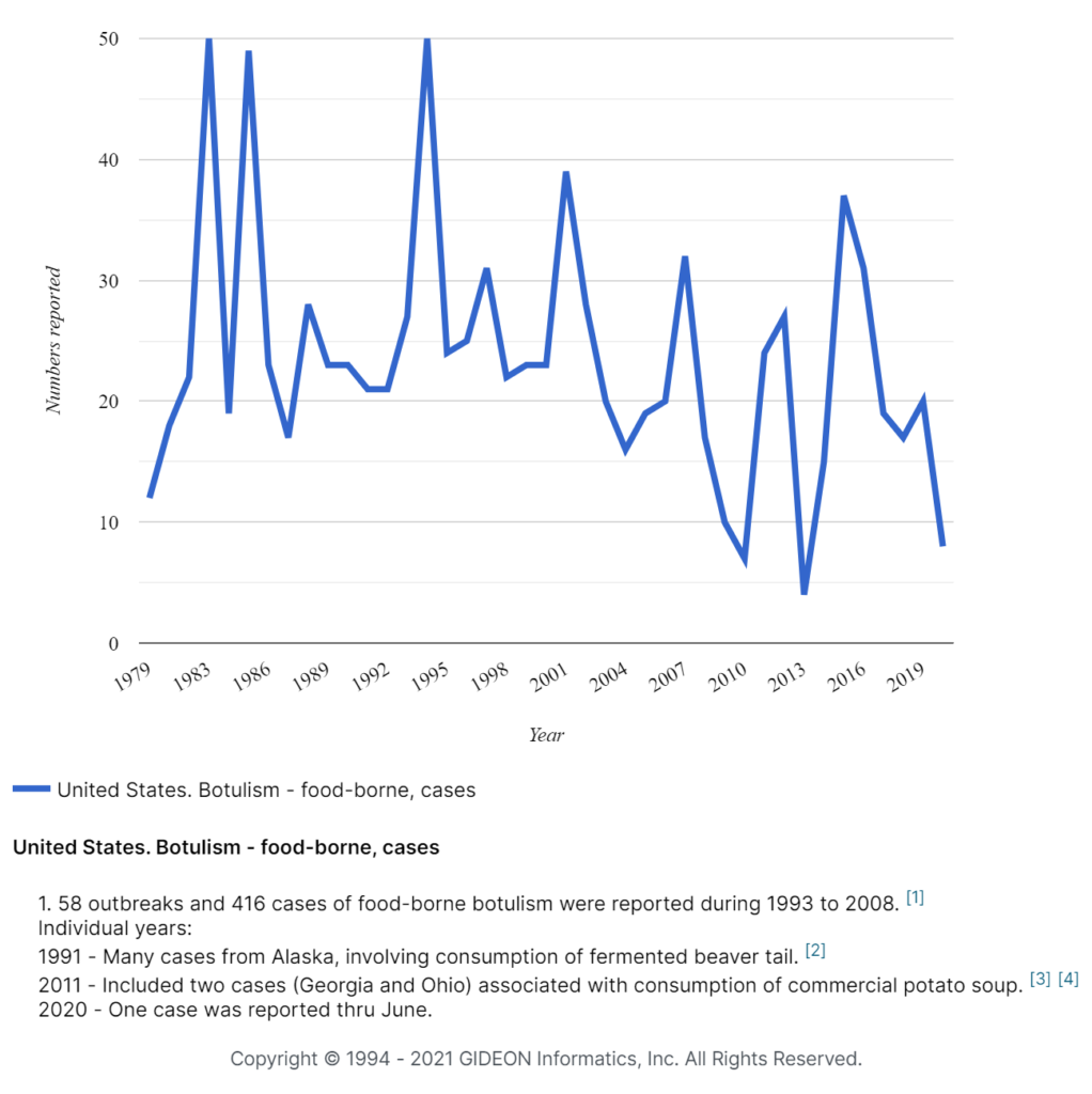
Botulism, a common and potentially fatal disease, is caused by Clostridium botulinum. Signs and symptoms usually appear within 18 to 36 hours of food ingestion Bacterial toxins can persist in the patient’s blood for up to 12 days. Common findings include respiratory failure and neurological symptoms, such as blurred vision, paralysis, etc. (2). 


Foodborne cases of botulism in the United States 1979-2019


Botulism cases in US, 1979 - 2020, GIDEON graph


Prevention and Management of Foodborne Illnesses

irradiated spinach_byMikeLichtContamination of food or food products can occur at various stages of production, including growth, processing, transportation, or storage. Thus, there is a critical need to adopt appropriate measures that correspond to these variable conditions to maximize food safety and minimize the transmission of foodborne illnesses.

 

1.     Decontamination of Fresh Produce

With rapid globalization, fresh produce is now available worldwide throughout the year in the form of frozen food.  Fresh produce must undergo proper decontamination before being frozen – to prevent such diseases as norovirus infection and hepatitis A (3, 4). Some of the common techniques used for decontamination include (5):

a.     Application of antibiotics during the growth stage

b.     Proactive sampling for detection of pathogens

c.     Bacteriocins (bacteria-generated toxins to kill the competitive strains)

d.     Antimicrobial natural products and nanoparticles

e.     Bacteriophages

f.      Irradiation 

Alternatively, consumers might receive probiotics and relevant vaccines to protect them from relevant foodborne illnesses.


2.     Development of Public Health Surveillance Systems

A competent public health surveillance system should be implemented by the Governmental and related organizations to identify impending outbreaks in order to facilitate effective policies and goals (6, 7). Such systems help control and prevent extensive transmission by collecting and analyzing epidemiological and clinical data that provide guidelines to take appropriate measures.


3.     Prophylactic Measures for People with Increased Susceptibility to Foodborne Illnesses

People who are vulnerable to foodborne infections, such as older adults, infants – and individuals with prior diseases of the immune system, liver, gastrointestinal tract, etc. should be particularly vigilant and observed preventive measures (8) including: 

a.   Adherence to a low-microbial diet and avoidance of undercooked meat, unpasteurized milk, etc.

b.  Consumption of  bottled natural water

c.   Infants should be preferably given sterile ready-to-eat formula; powdered formula should be reconstituted in boiling water and given in boiled water-  sterilized bottles

d.  Antimicrobial may be administered prophylactically during transplantation and other high-risk treatments.


4.     Food Safety Measures

In general, people should adopt safe food practices wherever they cook and consume food or food products (9). The cooking utensils/surfaces should be properly cleaned and sanitized. Raw and undercooked foods should be stored separately to avoid cross-contamination. Food should be cooked or reheated thoroughly, meats and seafood should be cooked to appropriate temperatures. Cooked food should be stored at < 5°C, and not for prolonged periods. Only clean, purified water should be used for cooking food to minimize any chances of waterborne infection. Raw fruits and vegetables should be properly washed before consumption.

Woman washing carrots and pomegranate in a metal sink
Raw fruits and vegetables should be properly washed before consumption.


At the Public Health level, comprehensive integration of government-issued guidelines and self-monitoring are needed to prevent and control foodborne illnesses.


Did you like this article? Share it on social media!

Check out more of our latest content here


References

  1.     “Foodborne diseases”, World Health Organization. [Online]. Available: https://www.who.int/health-topics/foodborne-diseases#tab=tab_1.
  2.     “Botulism”, GIDEON Informatics, Inc, 2021 [Online]. Available: https://app.gideononline.com/explore/diseases/botulism-10230.
  3.     Nasheri N, Vester A, Petronella N. Foodborne viral outbreaks associated with frozen produce. Epidemiol Infect. 2019 Oct;147:e291. doi: 10.1017/S0950268819001791.
  4.     Chapman B, Gunter C. Local Food Systems Food Safety Concerns. Microbiol Spectr. 2018 Apr; 6(2). doi: 10.1128/microbiolspec.PFS-0020-2017.
  5.     Yang SC, Lin CH, Aljuffali IA, Fang V. Current pathogenic Escherichia coli foodborne outbreak cases and therapy development. Arch. Microbiol. 2017 June; 199(6): 811-825. doi.org/10.1007/s00203-017-1393-y.
  6.     Ward H, Molesworth A, Holmes S, Sinka K. Public health: surveillance, infection prevention, and control. Handb Clin Neurol. 2018; 153:473-484. doi: 10.1016/B978-0-444-63945-5.00027-1.
  7.     Hoelzer K, Moreno Switt AI, Wiedmann M, Boor KJ. Emerging needs and opportunities in foodborne disease detection and prevention: From tools to people. Food Microbiol. 2018 Oct; 75:65-71. doi: 10.1016/j.fm.2017.07.006.
  8.     Lund BM, O’Brien SJ. The occurrence and prevention of foodborne disease in vulnerable people. Foodborne Pathog Dis. 2011 Sep; 8(9):961-73. doi: 10.1089/fpd.2011.0860.
  9.     “Five keys to safer food manual”, World Health Organization [Online]. Available: https://www.who.int/foodsafety/publications/consumer/manual_keys.pdf

Pathogen of the Month: Vibrio cholerae, the Causative Agent of Cholera

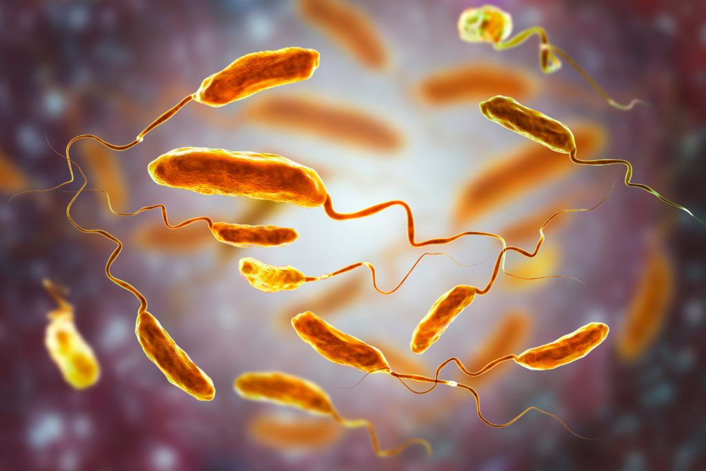
Vibrio cholerae
This blog was written by Dr. Jaclynn Moskow


Vibrio cholerae (V. cholerae) is a species of Gram-negative facultatively anaerobic bacteria of curved rod-shaped with single polar flagella. V. Cholerae has been classified into approximately 200 serogroups. Strains belonging to serogroups O1 and O139 cause the vast majority of cholera cases (1).

Vibrio cholerae is found naturally in brackish riverine, estuarine, and coastal waters. Recognized hosts of the organism include algae, shellfish, chironomid egg masses, fish, waterfowl, amebae, and copepods (2). V. cholerae colonies can form biofilms on both biotic and abiotic surfaces – including on shells, zooplankton, macroalgae, ship hulls, and plastic pollution (3-5). 


Cholera Transmission and Disease Severity 

Cholera is primarily transmitted through the consumption of fecally contaminated water and food. Foodborne outbreaks are most frequently linked to fish, shellfish, crabs, oysters, clams, rice, millet gruel, and vegetables (6).

Most V. cholerae infections are asymptomatic or mild in nature. Individuals with asymptomatic infections may still shed bacteria in their feces and infect others (7). Approximately 10% of V. cholerae infections will progress to severe disease (8). In endemic settings, the most severe infections occur in children, while in epidemic settings, severe disease occurs in adults as frequently as it does in children (9). 

Individuals with blood type O are more likely to suffer from severe V. cholerae infection (10). The use of drugs that reduce stomach acid, such as antacids, histamine receptor blockers, and proton pump inhibitors, also increases the risk of severe infection (11). 


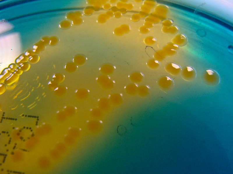
Vibrio cholerae bacteria on agar
Vibrio cholerae isolated from feces obtained from a patient with profuse diarrhea who had traveled to India. Photo courtesy of Nathan Reading


Signs and Symptoms of Cholera

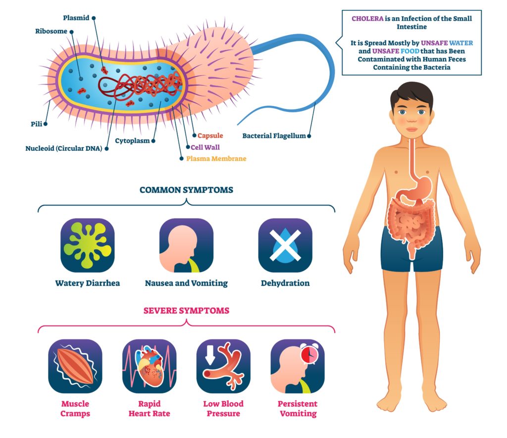
Cholera has an average incubation period of 1-5 days. Patients will experience a sudden onset of painless, watery diarrhea that may be accompanied by vomiting. The diarrhea is often characterized as having a “rice water” appearance and fishy odor. Fever is uncommon in adults, but often present in children (12).

In severe cases, dehydration may lead to the rapid progression to acidosis and electrolyte imbalance. Coma may occur. Without the replacement of fluids and electrolytes, hypovolemic shock and death ensue (12).

If left untreated, cholera has a 25-50% mortality rate. Proper treatment reduces the mortality rate to less than 1% (13).

Cholera vector illustration. Labeled infection structure and symptoms scheme. Educational infographic with unsafe water and food vibrio microorganism that causes diarrhea, vomiting and dehydration.

Diagnosis and Treatment of Cholera

Cholera is diagnosed via stool culture. When a case is suspected, healthcare and medical laboratory personnel should follow stool precautions.

Mild and moderate cases of cholera can be successfully treated with oral rehydration salts, while severe cases require rehydration with intravenous fluids (14). The World Health Organization (WHO) recommends reserving antibiotics as a treatment for severe cases only, as antibiotic use has no proven effect on controlling the spread of the disease and may contribute to antimicrobial resistance (15). In severe cases, tetracycline, doxycycline, azithromycin, erythromycin, or ciprofloxacin may be used (12). Most people who recover from V. cholerae infection incur long-lasting immunity (16).


Cholera Prevalence

Cholera originated in India and spread across the world during the 19th century (17). Since that time, there have been seven cholera pandemics, including one that is ongoing today (18). 

Currently, approximately 1.3 billion people are at risk for cholera in endemic countries. An estimated 2.86 million cholera cases occur annually, resulting in an estimated 95,000 deaths (19).

Over the last decade, the countries reporting the most cases of cholera have included Yemen, Somalia, the Democratic Republic of Congo, Mozambique, Bangladesh, and Haiti. If you have a GIDEON account, click here to explore the Cholera outbreak map. Cholera is exceedingly rare in Europe and the United States.


Cholera cases by region, 1953 – 2018

Cholera-cases-by-region-1953-2018


Cholera Prevention

Cholera cases by region, 1953 - 2018

When traveling to an area where cholera is endemic, precautions should include adherence to proper hand hygiene, drinking only bottled water, and avoiding uncooked food.

Ongoing worldwide efforts to end the current cholera pandemic center on increasing access to clean water and sanitation and expanding accessibility to existing cholera vaccines.


Did you like this article? Share it on social media!

Check out more of our latest content here


References 

(1) Morris, “Infections due to non-O1/O139 Vibrio cholerae”, Uptodate.com, 2019. [Online]. Available: https://www.uptodate.com/contents/infections-due-to-non-o1-o139-vibrio-cholerae

(2) “Cholera: Environmental Reservoirs and Impact on Disease Transmission”, One Health, pp. 149-165, 2014. Available: 10.1128/microbiolspec.oh-0003-2012

(3) B. Wucher, T. Bartlett, M. Hoyos, K. Papenfort, A. Persat and C. Nadell, “Vibrio cholerae filamentation promotes chitin surface attachment at the expense of competition in biofilms”, Proceedings of the National Academy of Sciences, vol. 116, no. 28, pp. 14216-14221, 2019. Available: 10.1073/pnas.1819016116

(4) C. Lutz, M. Erken, P. Noorian, S. Sun and D. McDougald, “Environmental reservoirs and mechanisms of persistence of Vibrio cholerae”, Frontiers in Microbiology, vol. 4, 2013. Available: 10.3389/fmicb.2013.00375 

(5) J. Moskow, “What Do Plastics Have To Do With Infectious Diseases and the Immune System?”, GIDEON Informatics, Inc, 2021. [Online]. Available: https://www.gideononline.com/2021/04/22/what-do-plastics-have-to-do-with-infectious-diseases-and-the-immune-system/ 

(6) G. Rabbani, W. Greenough. “Food as a vehicle of transmission of cholera”, J Diarrhoeal Dis Res, vol. 17, no. 1, pp. 1-9, 1999

(7) J. Lewnard, M. Antillón, G. Gonsalves, A. Miller, A. Ko and V. Pitzer, “Strategies to Prevent Cholera Introduction during International Personnel Deployments: A Computational Modeling Analysis Based on the 2010 Haiti Outbreak”, PLOS Medicine, vol. 13, no. 1, p. e1001947, 2016. Available: 10.1371/journal.pmed.1001947

(8) Cholera – Vibrio cholerae infection: General Information”, Centers for Disease Control and Prevention (CDC0, National Center for Emerging and Zoonotic Infectious Diseases (NCEZID), Division of Foodborne, Waterborne, and Environmental Diseases (DFWED), 2020. [Online] Available: https://www.cdc.gov/cholera/general/index.html

(9) J. Harris, R. LaRocque, F. Qadri, E. Ryan and S. Calderwood, “Cholera”, The Lancet, vol. 379, no. 9835, pp. 2466-2476, 2012. Available: 10.1016/s0140-6736(12)60436-x

(10) J. Harris and R. LaRocque, “Cholera and ABO Blood Group: Understanding an Ancient Association”, The American Journal of Tropical Medicine and Hygiene, vol. 95, no. 2, pp. 263-264, 2016. Available: 10.4269/ajtmh.16-0440

(11) S. Handa, “Which classes of medications increase the risk of cholera infection?”, Medscape.com, 2018. [Online]. Available: https://www.medscape.com/answers/962643-54708/which-classes-of-medications-increase-the-risk-of-cholera-infection 

(12) “Cholera”, GIDEON Informatics, Inc, 2021. [Online]. Available: https://app.gideononline.com/explore/diseases/cholera-10390

(13) J. Fournier and M. Quilici, “Choléra”, La Presse Médicale, vol. 36, no. 4, pp. 727-739, 2007. Available: 10.1016/j.lpm.2006.11.029 

(14) “WHO | WHO position paper on Oral Rehydration Salts to reduce mortality from cholera”, Who.int, 2021. [Online]. Available: https://www.who.int/cholera/technical/en/

(15) “Cholera”, Who.int, 2021. [Online]. Available: https://www.who.int/news-room/fact-sheets/detail/cholera 

(16) J. Harris, “Cholera: Immunity and Prospects in Vaccine Development”, The Journal of Infectious Diseases, vol. 218, no. 3, pp. S141-S146, 2018. Available: 10.1093/infdis/jiy414

(17) D. Lippi, E. Gotuzzo and S. Caini, “Cholera”, Paleomicrobiology of Humans, pp. 173-180, 2016. Available: 10.1128/microbiolspec.poh-0012-2015 

(18) S. Handa, “What are the 7 pandemics of cholera”, Medscape.com, 2018. [Online]. Available: https://www.medscape.com/answers/962643-54700/what-are-the-7-pandemics-of-cholera

(19) M. Ali, A. Nelson, A. Lopez and D. Sack, “Updated Global Burden of Cholera in Endemic Countries”, PLOS Neglected Tropical Diseases, vol. 9, no. 6, p. e0003832, 2015. Available: 10.1371/journal.pntd.0003832

Yellow Fever: Past and Present

1942, Innoculating eggs with yellow fever vaccine. USPHS (United States Public Health Service) Rocky Mountain Laboratory, Hamilton, Montana
Inoculating eggs with yellow fever vaccine, 1942. USPHS (the United States Public Health Service) Rocky Mountain Laboratory, Hamilton, Montana. Photographer: John Vachon


written by Dr. Jaclynn Moskow

What is Yellow Fever?

The yellow fever virus, a flavivirus, is transmitted via the bites of various mosquito species. The disease has an average incubation period of 3 to 6 days. The clinical presentation of yellow fever can vary greatly, ranging from a self-limited flu-like illness to overwhelming hemorrhagic fever – with a case fatality rate of 50%. Approximately 55% of yellow fever infections are asymptomatic, 33% are categorized as mild, and 12% severe (1).

Diagram of Symptoms of Yellow fever patient

Yellow fever generally manifests with the acute onset of fever, headache, backache, myalgia, and vomiting. Conjunctival infection may be seen, accompanied by facial flushing, relative bradycardia (Faget’s sign), and leukopenia. In some cases, fever and other symptoms may remit for a few hours to several days. Upon return of symptoms, icteric hepatitis and a hemorrhagic diathesis may follow with epistaxis, bleeding from the gums and gastrointestinal tract, and petechial and purpuric hemorrhages. Weakness, prostration, protracted vomiting, and albuminuria may also be noted. At this stage, patients will experience renal failure, myocardial dysfunction, necro-hemorrhagic pancreatitis, and seizures.

Yellow fever has a case fatality rate of 10 to 60% within 7 days of disease onset (1).


The Origin and Spread of Yellow Fever

Phylogenetic analyses indicate that yellow fever originated in Africa within the last 1,500 years. Its spread to the Americas coincided with the trans-Atlantic slave trade that began during the 16th century. It is likely that mosquitoes carrying yellow fever were imported into the Americas via slave ships (2). Significant outbreaks followed as the virus was introduced into populations with no pre-existing immunity. During the 18th and 19th centuries, approximately 25 major outbreaks claimed the lives of hundreds of thousands in New York City, Philadelphia, Baltimore, and New Orleans (3). Yellow fever also arrived in Europe during this time, with notable outbreaks occurring at Spanish, Portuguese, French, and British seaports (4).

It is estimated that for every soldier who died in battle during the Spanish-American War, 13 died of yellow fever (5). Yellow fever also killed many thousands during the construction of the Panama Canal. 

William C. Gorgas (1854-1920), on the site of Panama Canal construction.
William C. Gorgas (1854-1920), on the site of Panama Canal construction. As a U.S. Army surgeon during the Spanish American War, he established methods for eradicating mosquitoes hence reducing yellow fever and malaria among soldiers in Cuba. In 1904 he applied these techniques to control disease among the Panama Canal workers. Ca. 1910.


Development of a Vaccine for Yellow Fever

USA - CIRCA 1940: A stamp printed in USA shows portrait of Dr. Walter Reed, series Scientists, circa 1940

Early attempts to develop a vaccine for yellow fever resulted in the deaths of several test subjects and researchers. In 1900, a team led by Major Walter Reed traveled to Cuba to study the disease. At this point, the medical community was largely dismissive of the theory that mosquitos were the vectors of transmission for yellow fever. Working under the assumption that the mosquito theory was indeed incorrect, Reed’s team began experimenting with mosquitos and volunteers. After receiving criticism about using human test subjects, some team members decided to instead experiment on themselves. Unfortunately, this resulted in the death of physician-scientist Dr. Jesse Lazear – but with his death, the mosquito theory began to gain acceptance (6). 

Despite the death of Dr. Lazear, yellow fever research on human test subjects continued. The Reed team conducted a second and third set of mosquito experiments, offering financial compensation in the form of gold to study participants (6). After learning that none of these latest Reed study participants had died, Cuban physician Dr. John Guiteras began his own experiments. Unfortunately, three of his 42 test subjects succumbed to the illness, and with this, yellow fever research in Cuba came to a halt (7).

Subsequent efforts to control yellow fever centered on reducing mosquito populations as opposed to vaccine development, until 1918 when the Rockefeller Foundation began conducting yellow fever research. Within a year, Japanese scientist Dr. Hideyo Noguchi, who was working with the Foundation, announced that he had successfully developed a vaccine. Individuals in the United States, Latin America, and the French African colonies began receiving his vaccine, but the legitimacy of the studies leading to its development were soon called into question, and ultimately the vaccine was pulled (7).

The Rockefeller Foundation continued its efforts, and in 1925, they sent investigators to Lagos to determine if the African and South American diseases were caused by the same pathogen. Unfortunately, this trip resulted in three more investigators contracting and dying from yellow fever, including Dr. Noguchi. Nonetheless, the Rockefeller Foundation persisted, and a few years later, another candidate vaccine was developed – this time from efforts led by Dr. Max Theiler (7).

Despite the history of physician deaths related to yellow fever experimentation, Brazilian physician Dr. Bruce Wilson volunteered to receive the first dose of the Theiler vaccine. It was designated a success, and mass production began. Soon after, the Pasteur Institute developed their own vaccine, and for the next several years, the Rockefeller Foundation vaccine was used in the West as well as in England, and the Pasteur Institute vaccine used in France and its African colonies (7).

During World War II, the Rockefeller Foundation vaccine was given to almost all US soldiers. Unfortunately, the vaccine contained blood serum, and vaccination efforts resulted in approximately 330,000 soldiers contracting hepatitis B virus infection (8). Blood serum was subsequently removed as a component of the vaccine, and in 1953 a yellow fever vaccine was licensed for civilian use in the US (9). Use of the Pasteur Institute vaccine eventually ceased due to cases of postvaccinal encephalitis, but a variant of the Rockefeller foundation vaccine is still used today. Dr. Theiler received the Nobel Prize in Physiology or Medicine for his critical role in its development.


Yellow Fever in 2021

Yellow fever vaccination est. coverage 1980 - 2019

Today, the Centers for Disease Control and Prevention (CDC) recommends vaccination against yellow fever for individuals 9 months and older and who are traveling to or living in areas at risk in Africa and South America (10). It is a live attenuated vaccine, and thus contraindicated in patients who are immunocompromised. The vaccine is highly effective, with a median seroconversion rate of 99% (range 81–100%) in clinical trials (11).

Yellow fever is currently estimated to affect 200,000 people each year, resulting in 30,000 deaths, with 90% of cases occurring in Africa (12). Recent outbreaks have occurred in Brazil, Angola, Nigeria, and the Democratic Rep. of Congo. If you have a GIDEON account, click here to explore the Yellow Fever outbreak map. There are ongoing efforts to expand accessibility to the vaccine in these regions, as well as to implement additional vector control programs. 

The last outbreak of Yellow Fever in the United States occurred in 1905 (13). Yellow fever outbreaks ceased in Europe after World War II, when, for unknown reasons, the Aedes aegypti mosquito disappeared (14).

The absence of yellow fever in Asia is not fully understood. Some have speculated that differences in mosquito species may play a role. Another theory is that there may be a cross-immunity between yellow fever and other flaviviruses endemic to Asia, such as dengue fever. A third theory is that yellow fever has simply never been introduced into Asia (2).


Did you like this article? Share it on social media!

Check out more of our latest content here


References

(1) “Yellow fever”, GIDEON Informatics, Inc, 2021. [Online]. Available: https://app.gideononline.com/explore/diseases/yellow-fever-12650

(2) Cathey and J. Marr, “Yellow fever, Asia and the East African slave trade”, Transactions of the Royal Society of Tropical Medicine and Hygiene, vol. 108, no. 5, pp. 252-257, 2014. Available: 10.1093/trstmh/tru043 

(3) F. Douam and A. Ploss, “Yellow Fever Virus: Knowledge Gaps Impeding the Fight Against an Old Foe”, Trends in Microbiology, vol. 26, no. 11, pp. 913-928, 2018. Available: 10.1016/j.tim.2018.05.012 

(4) M. Morillon, B. Marfart, and T. Matton, “Yellow fever in Europe in 19th Century”, Ecological Aspects of Past Settlement in Europe. P. Bennike, pp. 211-222, 2002.

(5) Staples, “Yellow Fever: 100 Years of Discovery”, JAMA, vol. 300, no. 8, p. 960, 2008. Available: 10.1001/jama.300.8.960 

(6) “Politics of Participation: Walter Reed’s Yellow-Fever Experiments”, AMA Journal of Ethics, vol. 11, no. 4, pp. 326-330, 2009. Available: 10.1001/virtualmentor.2009.11.4.mhst1-0904

(7) J. Frierson. “The yellow fever vaccine: a history”, Yale J Biol Med, vol. 83, no. 2, pp. 77-85, 2010

(8) M. Furmanski. “Unlicensed vaccines and bioweapon defense in World War II”, JAMA, vol. 282, no. 9, p. 822, 1999

(9) “Historic Dates and Events Related to Vaccines and Immunization”, Immunize.org, 2021. [Online]. Available: https://www.immunize.org/timeline/

(10)”Yellow Fever Vaccine Recommendations”, Centers for Disease Control and Prevention, National Center for Emerging and Zoonotic Infectious Diseases (NCEZID), Division of Vector-Borne Diseases (DVBD), 2021. [Online]. Available: https://www.cdc.gov/yellowfever/vaccine/vaccine-recommendations.html

(11) J. Staples, A. Barrett, A. Wilder-Smith and J. Hombach, “Review of data and knowledge gaps regarding yellow fever vaccine-induced immunity and duration of protection”, npj Vaccines, vol. 5, no. 1, 2020. Available: 10.1038/s41541-020-0205-6 

(12) “Yellow Fever”, Centers for Disease Control and Prevention, Global Health, 2018. [Online]. Available: https://www.cdc.gov/globalhealth/newsroom/topics/yellowfever/index.html

(13) K. Patterson, “Yellow fever epidemics and mortality in the United States, 1693–1905”, Social Science & Medicine, vol. 34, no. 8, pp. 855-865, 1992. Available: 10.1016/0277-9536(92)90255-o 

(14) “Facts about yellow fever”, European Centre for Disease Prevention and Control, 2021. [Online]. Available: https://www.ecdc.europa.eu/en/yellow-fever/facts


GIDEON Sign In