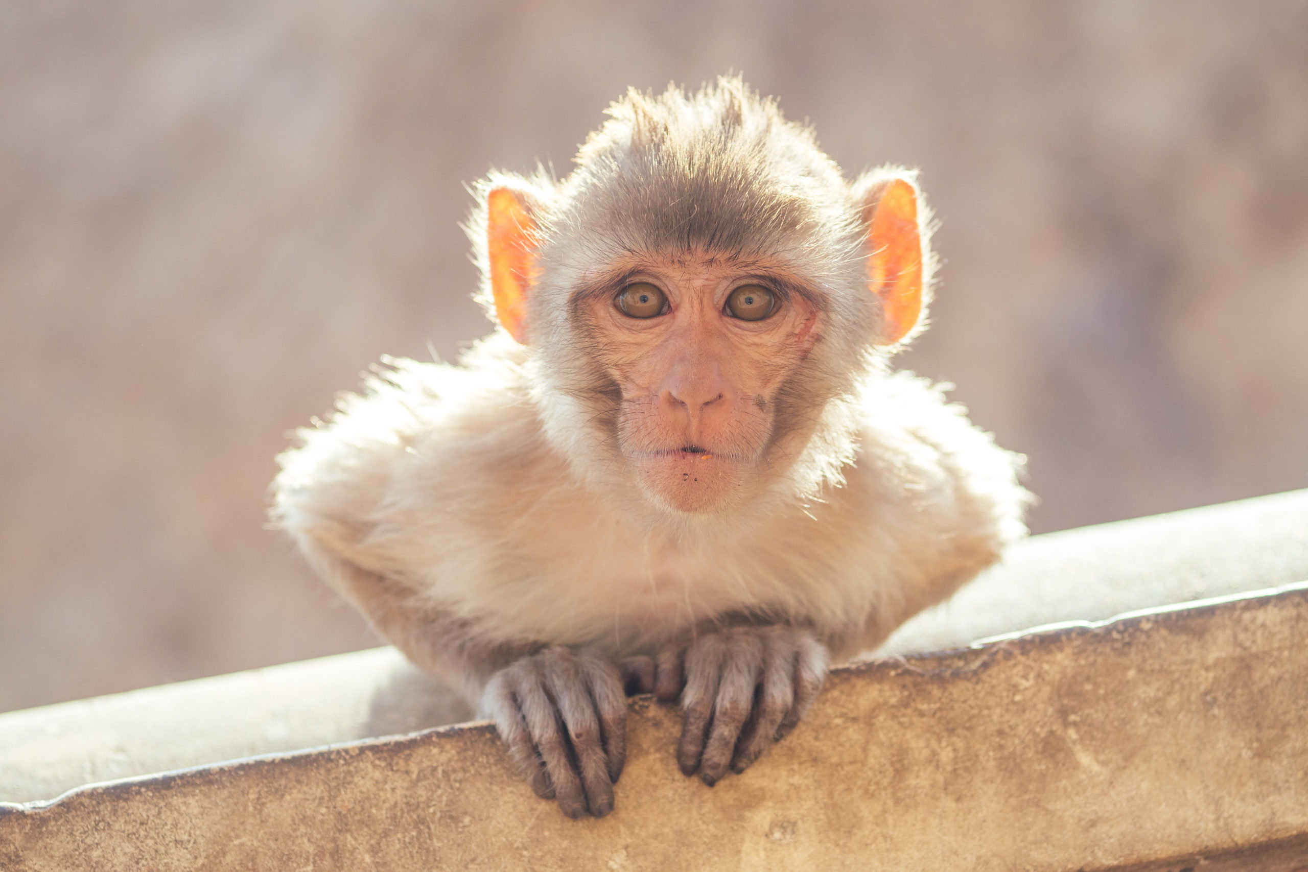
Cynomolgus monkey, a known reservoir of the Monkeypox virus
WHAT IS MONKEYPOX?
Monkeypox, as the name implies, is a disease of monkeys (unlike chickenpox – which has no relation to chickens). Although the condition is reported in a group of eleven African countries, the virus was first discovered in a laboratory in Denmark in 1958, when it was first isolated from cynomolgus monkeys.
The signs and symptoms are similar to those of smallpox. Following a three-day prodrome of fever, headache, myalgia, and back pain, patients develop a papular rash in the face, extremities, and genitals. The rash then spreads outward to involve the face, with lesions evolving into umbilicated pustules.
Unlike smallpox, death from monkeypox is relatively uncommon – five-to-ten percent.
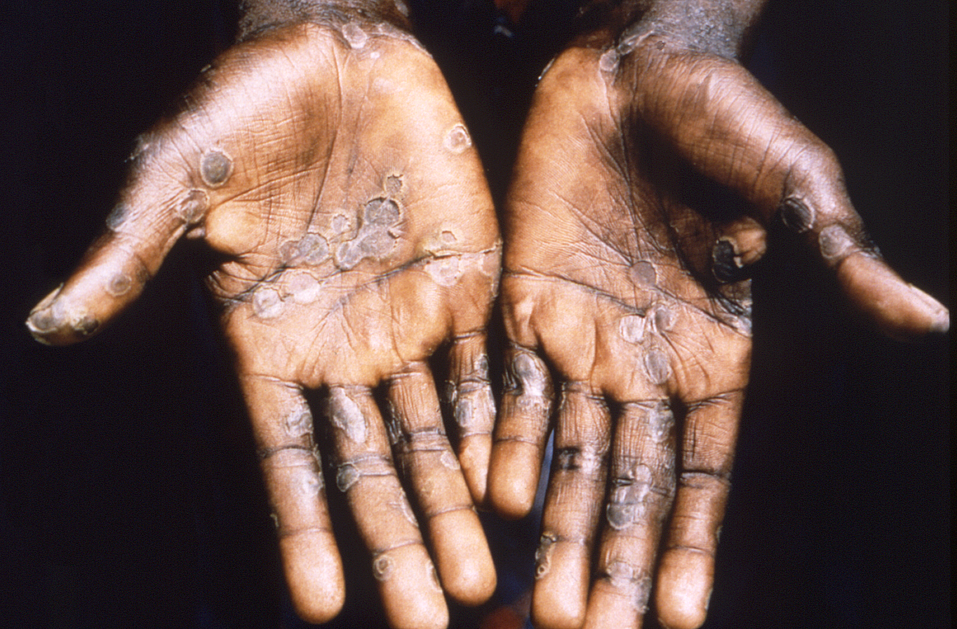
This 1997 image was created during an investigation into an outbreak in the Democratic Republic of the Congo (DRC) and depicts the palms of a monkeypox case-patient. It is important to note how similar this maculopapular rash appears to be when compared to the rash of smallpox, also an Orthopoxvirus. Image courtesy of CDC/Dr. Brian W.J.Mahy
NOTABLE OUTBREAKS
During the single year of 1967, almost eleven thousand cases occurred in West and Central Africa.
The most unusual outbreak of monkeypox occurred in 2003 when 81 humans in the American Midwest were infected through contact with infected prairie dogs – themselves infected by rodents that had been imported from Ghana. Fortunately, all patients recovered without sequelae. By coincidence, the iconic outbreak of SARS was also reported at this time – perhaps, as in the current COVID-19 pandemic, distracting media attention from events surrounding other diseases.
During 2018 to 2019, five Nigerian travelers were found to have monkeypox – in Israel, Singapore, and London.
A RECENT RISE IN CASES
The viruses of monkeypox and smallpox are biologically similar. Indeed an attack of one will immunize the patient against the other. Thus, rates of monkeypox were low during the period that smallpox vaccination was widely used in Africa, while the discontinuation of vaccination has been followed by a resurgence of monkeypox cases.
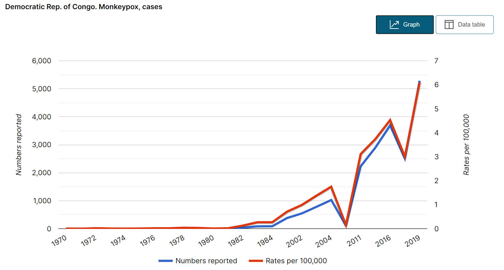
These developments are well illustrated by an ongoing outbreak of monkeypox that has persisted well into the COVID-19 pandemic. From January 1 to September 13, a total of 4,494 cases of monkeypox (171 fatal) were reported in the Democratic Republic of Congo.
Cases in the Democratic Republic of Congo, 1970 – 2019
Did you like this article? Share it on social media!
What is the difference between antigen vs antibody, and what role do they play in creating an effective vaccine? With the recent focus on the development of a COVID-19 vaccine there has been much talk of antigens and antibodies, often interchangeably, and little clarity on what they are – or the role they play in creating an effective vaccine. In this blog, we’ll cut through the jargon and discover the facts together.
Antigen
An antigen is any substance or organism that is unrecognized by our immune system. It could be anything from bacteria to chemicals, to viruses … or even foods [1]. Antigens typically trigger an immune response, which may consist of an antibody (more on that later), and are classified by their origins [2]:
Exogenous: entering from outside the body
Endogenous: generated from within
Autoantigens: proteins targeted in autoimmune diseases
Neoantigens (or tumor antigens): resulting from tumor cells.
Native antigens: An antigen which will later be processed by an antigen-presenting cell
In some cases, these main types have subtypes – but we won’t get into an immunology lecture today. An antigen-presenting cell is a cell that processes and then presents the antigen to T-cells (a form of white blood cells), which can then ‘handle’ the antigen, often by killing the offending cell [3].
Your immune system has “memory” which allows the system to deal with the offending antigen much more quickly and efficiently the next time it is encountered. Vaccines are designed to simulate that first encounter with an antigen and create a robust memory in case the offending agent reappears in the future. [4].
The importance of vaccines is covered in more detail here, but in short, antigens themselves are crucial in the development of vaccines. Generally, the vaccine consists of a potentially hostile antigen, in a very weak or inactive form.
Antibody
Antibodies are proteins that bind with the antigen in order to neutralize the latter – or make other elements of the immune system “aware” of their presence. Antibody-producing cells are specifically designed to tackle one type of antigen; and your blood, bone marrow, lymph glands, and spleen will contain millions of them to ensure that every known antigen will be confronted by a corresponding antibody [5].
Antibodies are secreted by B leukocytes (a form of white blood cell) and circulate in blood plasma either freely or attached to the surface of a B cell. The B and T cells work in unison to identify and locate antigens, create the correct antibodies, and capture (kill/neutralize) the antigen [6].
A vaccine, by exposing the immune system to a new antigen, will “teach” antibodies the correct format in which to capture or tag that antigen. When the actual disease antigen later enters the body, the immune system will rapidly respond with minimal discomfort and inconvenience.
Effective vaccination needs both
To summarize – an antigen is a disease agent (virus, toxin, bacterium parasite, fungus, chemical, etc) that the body needs to remove, and an antibody is a protein that binds to the antigen to allow our immune system to identify and deal with it.
Antigens and antibodies work in tandem when vaccinating
Don’t take this all for granted, though. As impressive as our immune system is, it’s far from perfect and needs our assistance to prevent harmful antigens from entering the body – through hand washing, face masks, and social distancing. Look after your body and it will look after you!
Did you like this article? Share it on social media!
[3] T. Kambayashi and T. Laufer, “Atypical MHC class II-expressing antigen-presenting cells: can anything replace a dendritic cell?”, Nature Reviews Immunology, vol. 14, no. 11, pp. 719-730, 2014. Available: 10.1038/nri3754
[4] A. Abbas, A. Lichtman and S. Pillai, Cellular and molecular immunology, 9th ed. Philadelphia: Elsevier, 2018, p. 97.
[5] C. Janeway, Immunobiology 5: the immune system in health and disease, 5th ed. Garland Publishing.
[6] L. Borghesi and C. Milcarek, “From B Cell to Plasma Cell: Regulation of V(D)J Recombination and Antibody Secretion”, Immunologic Research, vol. 36, no. 1-3, pp. 27-32, 2006. Available: 10.1385/ir:36:1:27
posted inExamples, News, Therapy | Comments Off on Antigen vs antibody – what is the difference?
Dog vaccination programs are the most effective way to prevent Rabies
Rabies is endemic to over 150 countries, and according to the World Health Organization, 99% of all transmissions to humans are from dogs, potentially bringing into question the animal’s status as the ‘man’s best friend’.
In Europe, southern Africa, and parts of North America, most cases are acquired from wild carnivores; mongooses, and vampire bats in Latin America and the Caribbean. In more recent years, humans have acquired rabies from inhalation of aerosols in bat caves, ingestion of dogs and cats for food, ticks, cart-scratches, and inadvertent transplantation of corneas or internal organs from infected donors.
In recognition of World Rabies Day, we have asked our co-founder, Dr. Stephen Berger, for his take on the disease. He didn’t hold back with the assessment!
“Rabies, from an evolutionary standpoint, is a truly “stupid” disease. Most animals with Rabies virus infection become paralyzed and die – thereby preventing the survival and reproduction of the virus itself. Ebola and Smallpox, albeit highly contagious, are also “stupid” in this respect,” said Dr. Berger, highlighting an interesting point.
When a disease limits its access to new hosts, how does it survive and continue to spread?
How long before it’s too late?
Virus transmission takes place via exposure to infected saliva, not necessarily a bite – although bites are the most common means of transmission. The virus is very active in moist conditions but quickly becomes non-infectious when dried or exposed to direct sunlight.
In dogs, the virus incubates for between 2 weeks to 4 months before the animal can transmit the disease.
In human cases, the incubation period can be as short as a few days or take as long as a few months for the disease to become active and symptoms to show. In rare cases, Rabies has appeared as long as five years following an animal bite. Once symptoms appear, the case-fatality rate is virtually 100%. As of 2014, only 13 cases of human survival from rabies had been documented.
Two forms of Rabies
The disease has two recognized forms – Furious and Paralytic, also known as ‘dumb Rabies’. In the furious form, dogs (or humans) froth at the mouth and display extreme hyperactivity. Patients often develop spasms when exposed to water – thus the term “hydrophobia.” In some cases, a similar response will follow fanning the patient – “aerophobia.”
While Furious Rabies is most familiar to the lay public (thanks to the cinema and TV) the Paralytic form often predominates among dogs. This type causes a slow progression from difficulty swallowing to full-body paralysis and eventually, death.
Rabies is 100% vaccine-preventable
When compared to diseases such as Tuberculosis, where symptoms such as coughing actively support transmission through the projection of infectious material into the air, Rabies would seem easier to control, especially when effective preventative and therapeutic vaccines and immune-globulins are available.
The disease was recorded as far back as 556 BC, in China, and a viable vaccine has existed since 1885. The World Health Organization (WHO) considers the disease to be one of the Neglected Tropical Diseases, in part due to the under-usage of effective vaccination, which works even after the virus has entered the body. A lesser-known adjunct to therapy is vitally important, but not as well-known to the public. Thorough cleansing of a wound using soap and water has been shown to reduce the incidence of Rabies by 50% following the bite of a rabid animal!
Sadly, despite the fact that 29 million people receive the vaccine every year, thousands still die from the disease. 95% of deaths occur in Asia and Africa, with children under the age of 15 making up 40% of all cases.
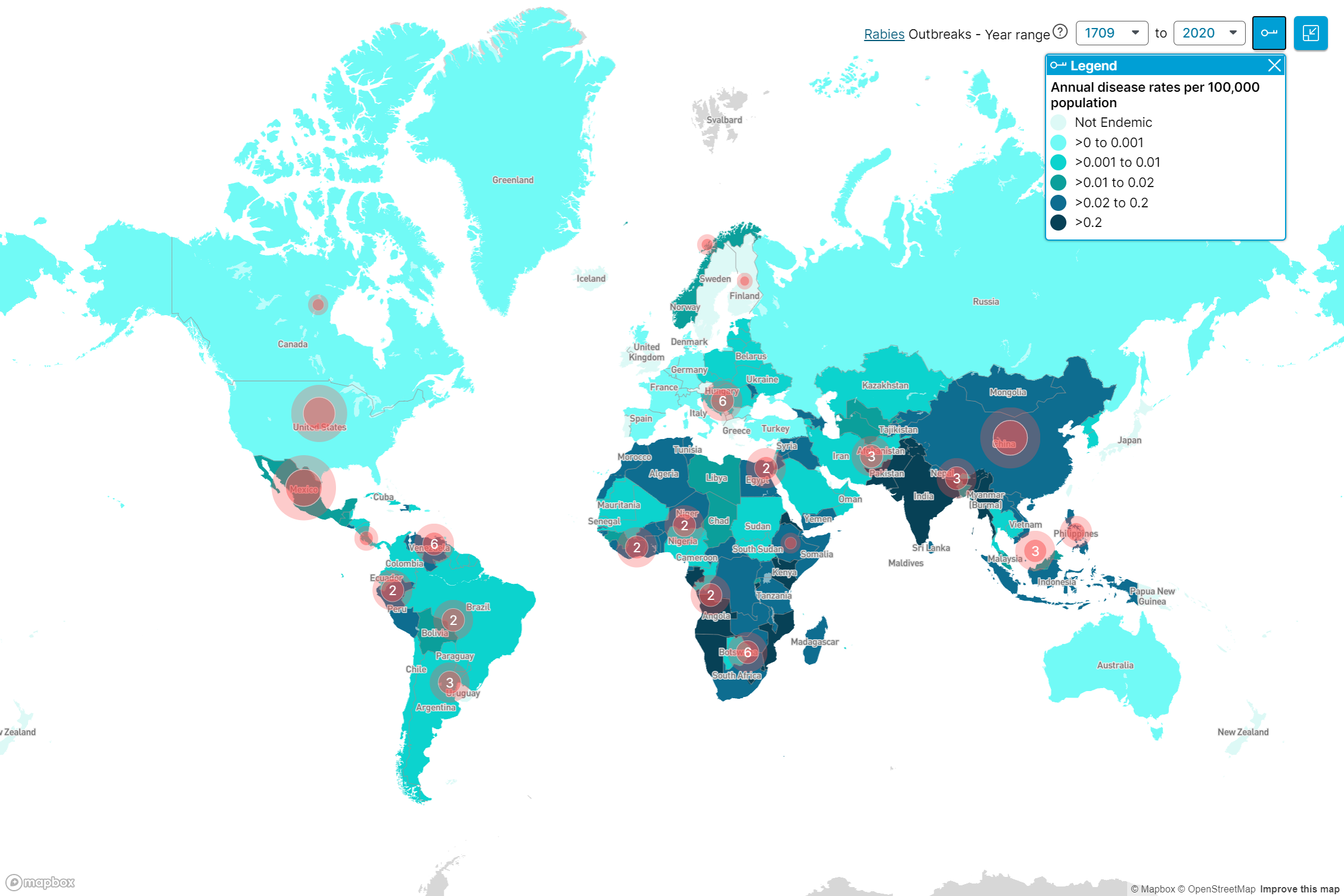
Stray dogs present a significant problem in countries such as India, Namibia, Angola, Mozambique, and Kenya, where annual disease rates exceed 0.2 per 100,000 population.
WHO leads a collective “United Against Rabies” campaign to drive progress towards zero human deaths from dog-mediated Rabies by 2030.
Rabies distribution and outbreaks map, 1709 – 2020
Did you like this article? Share it on social media!
There have been many happy accidents in science. Several of these were of great benefit to medicine.
For example, in 1895, a German physicist working with a cathode ray tube happened to place his hand in front of the rays and found that he could see his bones in the image projected onto the screen. Soon after that, the first X-ray images were produced.
There have been other instances where serendipity played a role in unearthing effective treatments against diseases.
THE FIND OF THE 20TH CENTURY
The most famous of these happy accidents is the discovery of Penicillin as an antibiotic remedy.
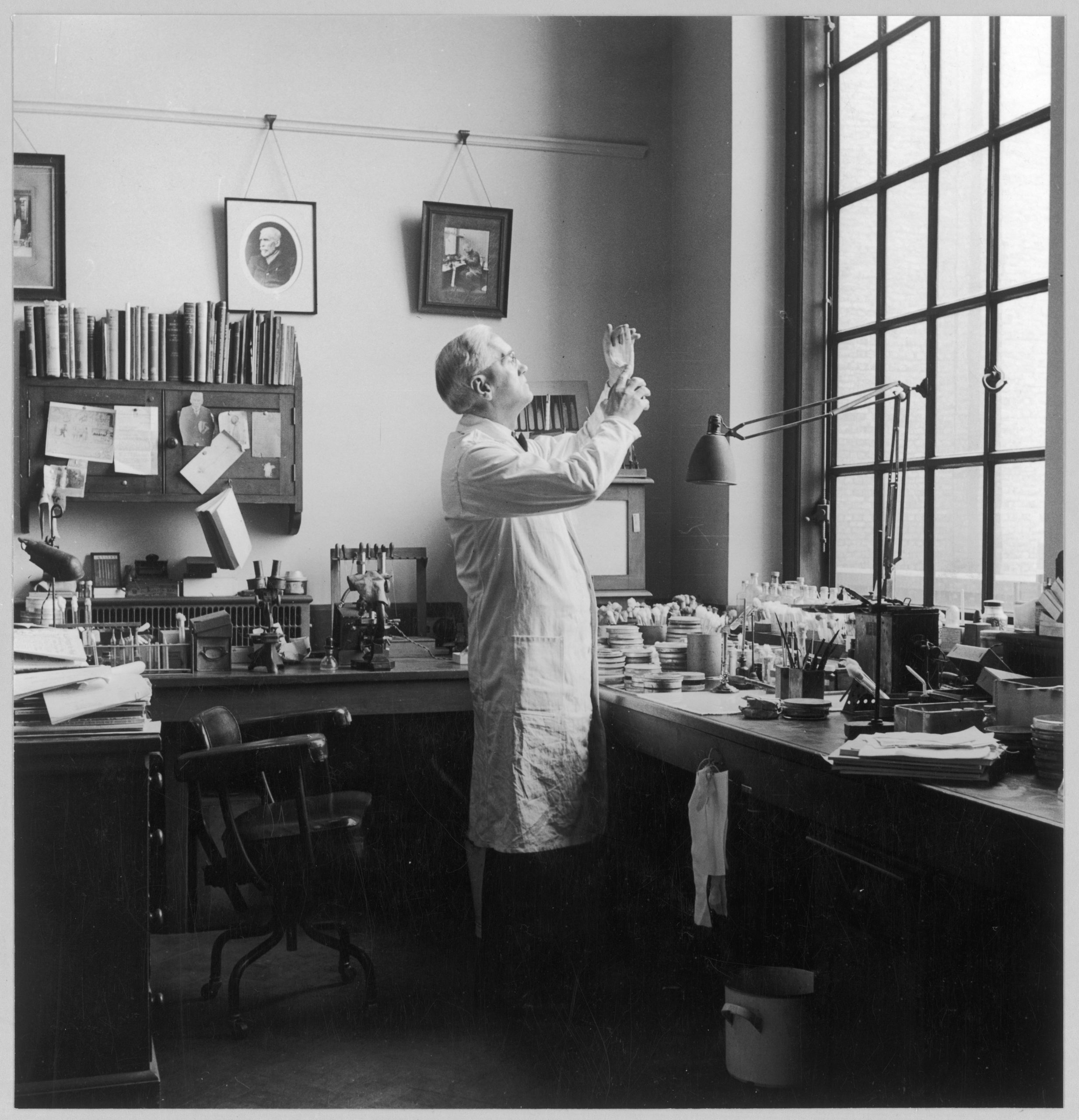
Alexander Fleming, a Scottish bacteriologist, worked at the inoculations department at St Mary’s Hospital in the early 1900s.
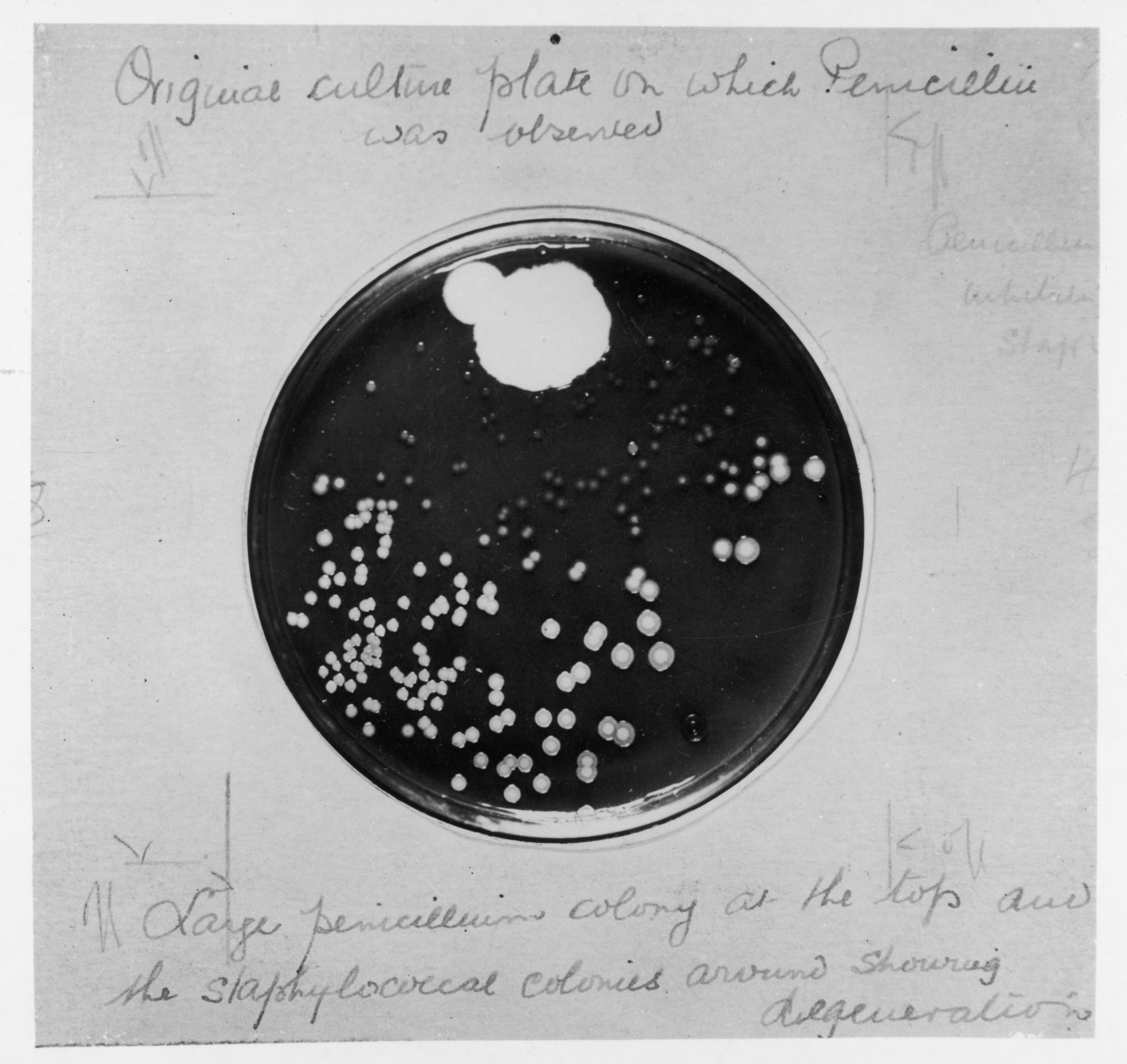
In September 1928, Fleming had left a pile of bacteria cultures in his laboratory before going on holiday with his family. The cultures he was studying were known to cause septic infections. By accident, he left one of the Petri dishes uncovered.
Fleming returned to find that a bluish-green mold, similar to the mold found on bread, had contaminated the specimen. The area around the mold in the Petri dish was clear of bacteria.
Fleming observed that the mold seemed to have killed the germs. This mold was identified as a strain of Penicillium. He saw this as a potential treatment for bacterial infections.
Penicillin culture, 1929
IMPORTANCE OF SHARED SCIENCE
Fleming was able to further identify that it wasn’t just the mold that killed the bacteria but the ‘juice’ the mold seemed to produce.
He also discovered that the ‘mold juice‘ was effective against pathogens that are responsible for diseases like Meningitis, Diphtheria and Gonorrhea.
Fleming’s effort would bear no further fruits. He was not able to produce and purify the ‘mold juice’ in substantial quantities.
However, he named the substance Penicillin and published his findings in the British Journal of Experimental Pathology in 1929. This crucial step allowed others to build on his work.
A decade later, Fleming’s findings piqued the interest of two Oxford scientists: Howard Florey and Ernst Chain. Eventually, they found a way to mass-produce the antibiotic in a form that could kill harmful bacteria without having any toxic effects on the human body.
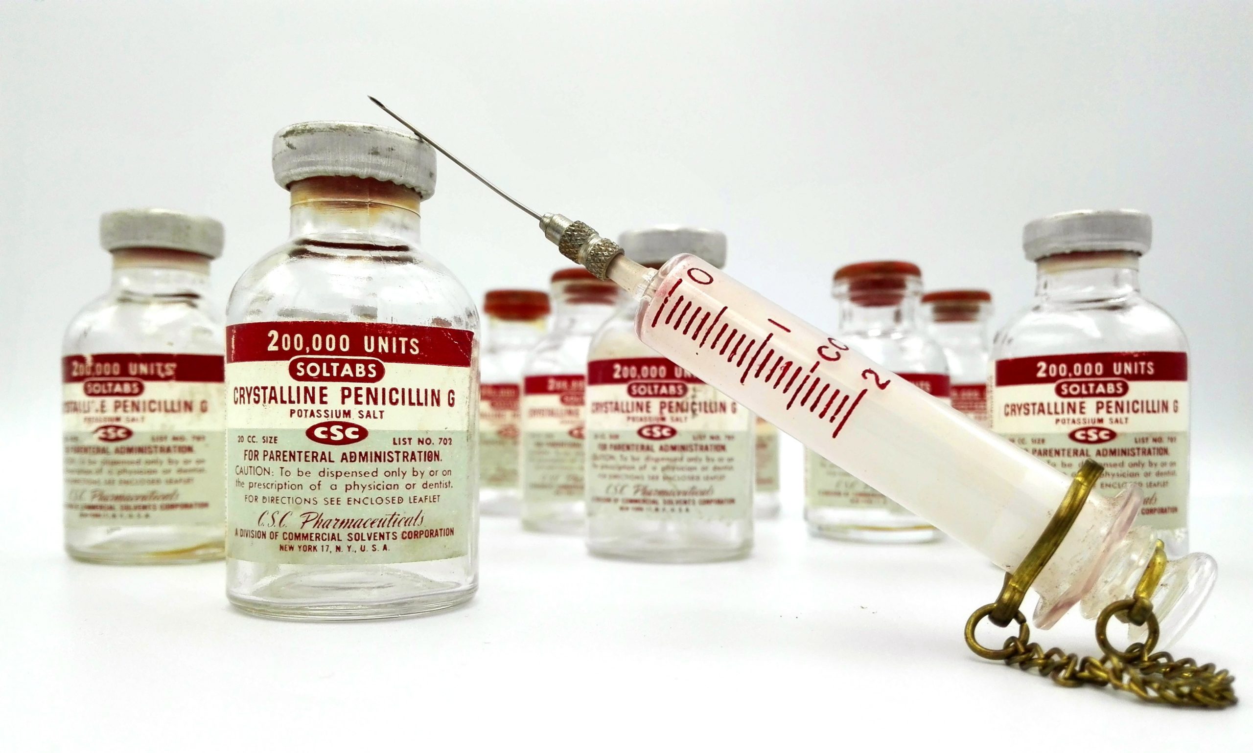
Vintage vials of Penicillin G
PENICILLIN’S WARTIME VALUE
During World War I, Alexander Fleming was stationed in France and served in the Army Medical Corps as a captain. He observed that the death of soldiers was not always from wounds inflicted in battle, but rather from bacterial infections.
The principal treatment of such infections consisted of the administration of antiseptics. Fleming noted that these often did more harm than good. He wrote about this, however, his findings were not taken seriously at the time.
During World War I, the death rate from bacterial pneumonia was 18%. In WWII, thanks to Penicillin, the death rate from the same condition fell to less than 1%. This enabled many soldiers to return home in good health.
AN EXCEPTIONAL DISCOVERY
The mass production of Penicillin is credited with saving the lives of many thousands of soldiers during World War II.
Antibiotics of the Penicillin family have been found to cure a wide variety of bacterial infections from mild, moderate upper respiratory tract infections to skin ulcers and urinary tract infections.
In 1944, Alexander Fleming was knighted by King George VI. In 1945, he received a Nobel Prize in Physiology or Medicine, together with Howard Florey and Ernst Boris Chain.
The praise was well deserved, as infections that were once life-threatening are now only mild inconveniences because of Penicillin’s versatility and efficacy. Penicillin richly deserves its place as one of the most important anti-infective drugs of all time.
Interestingly, Fleming was not the first to observe the antibacterial effect of Penicillium. Between 1868 and 1873, a famous surgeon named Theodor Billroth discovered that it inhibited bacterial growth – but nothing was done about it at the time. He died when Fleming was 13 years old.
Did you like this article? Share it on social media!
Brucellosis is most frequently transmitted via unpasteurized dairy products
Zoonotic diseases seem to be keeping the world on its toes. What is the disease responsible for the latest outbreak in China and what is its pathogenic potential?
Not the next COVID-19
Brucellosis is a category B bioterror disease, as classed by CDC. While it is one of the most important zoonotic diseases worldwide, brucellosis has limited pandemic potential, since human-to-human transmission is sporadic and occurs via blood, sexual exposure, or breastfeeding.
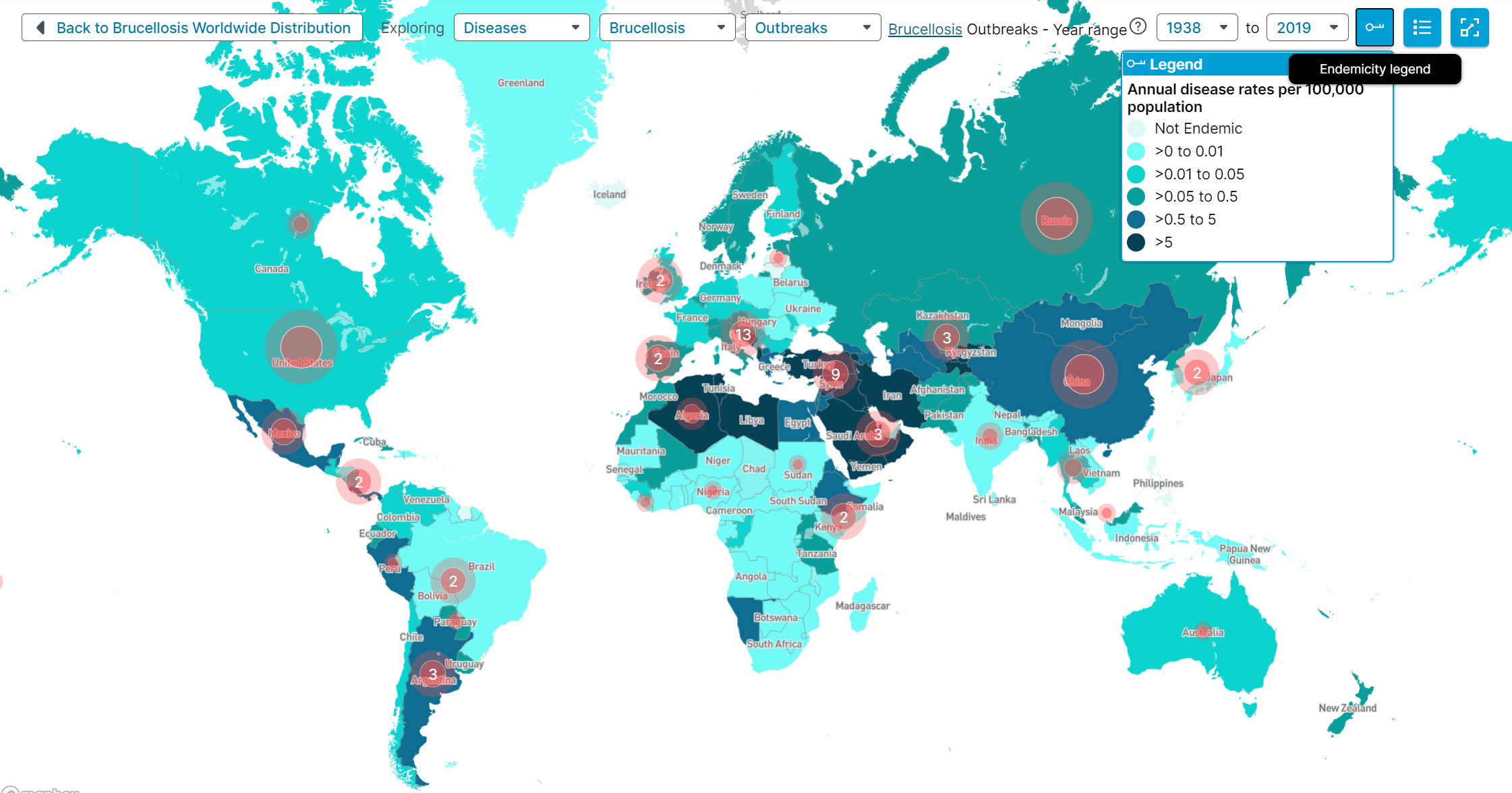
63% of cross-border events since 1965 were directly linked to the consumption of unpasteurized dairy products. The largest ever reported outbreak took place in the province of Ghardaia, Algeria, in 2016. During that time, 819 cases were recorded – health authorities suspected consumption of raw milk and a popular traditional cheese “Kamaria” may have been to blame. Epizootics (outbreaks among animals) can be much larger. Over 40,000 cattle acquired the disease during an outbreak in Spain in 2010.
Brucellosis outbreaks and distribution map, 1938 – 2019
What are the symptoms of Brucellosis?
Initial symptoms include fever, sweats, and pain in muscles and joints; while protracted infections may involve the heart valves, liver, or testicles.
Occupational hazard
The outbreak in China occurred among biopharmaceutical plant workers; and several prior disease clusters have involved workers in hospital laboratories. For this reason, individuals working with Brucella must be especially careful when handling this pathogen.
For instance, in 2007, a biodefence laboratory in the United States was closed after workers were exposed to two bioterror agents: Brucella (agent of Brucellosis) and Coxiella burnetii (agent of Q fever). Fortunately, this incident did not result in an actual outbreak. Professionals working in such environments are well-prepared for the possibility of similar scenarios and will likely behave in a way that minimizes any risks to public health.
Interested in learning more? Check out our ebook Brucellosis: Global Status for the latest epidemiological data, clinical findings, and potential use in bioterrorism. The ebook includes 175 graphs and 1,977 references.
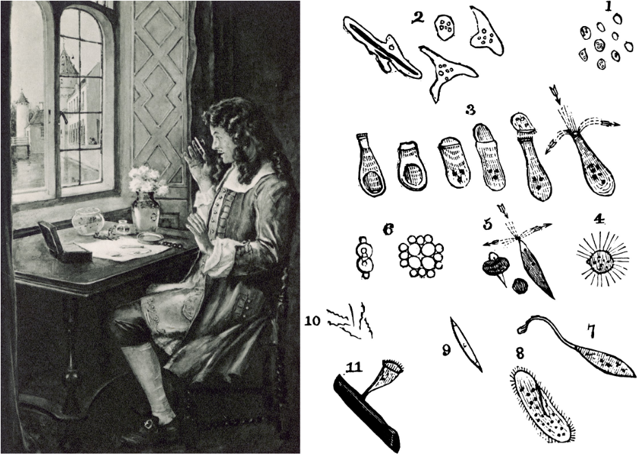
Antonie van Leeuwenhoek and a drawing of animalcules
Some discover their aptitude for science by natural curiosity, which causes them to investigate their surroundings. In doing so they find many hidden secrets that only curiosity like theirs could have revealed. However, an inquisitive nature alone doesn’t make one a scientist. Explorers, adventurers, reporters, and criminal investigators all lead lives based on it too.
Something special happens when curiosity is coupled with an empirical mind. That combination begins to approach the scientific method. The only thing left is to provide a record of findings so that other scientists can attempt to falsify the results.
Scientist By Nature
Antonie van Leeuwenhoek did all of this and more. He used the scientific method to unearth the existence of previously unseen organisms, and he was in regular correspondence with the Royal Society in London, discussing his findings.
Leeuwenhoek was a businessman by trade, but a scientist by nature. His skill in grinding glass allowed him to produce single-lens microscopes that could magnify over 200 times.
On 17th September 1683, Leeuwenhoek was the first to report the existence of bacteria seen through his microscopes. He called them little “animalcules”.
He achieved clearer and brighter images than any of his scientific fellows would achieve for centuries. This led to doubts and questions about the certainty of what he claimed to have seen.
It wasn’t until 1981 that Leeuwenhoek’s original specimens at the Royal Society were successfully photographed. Even this was done using one of his surviving microscopes. This finally dispelled the lingering disbelief that he indeed saw what he claimed.
However, Leeuwenhoek’s greatest delights and findings were in the field of microbiology. His discoveries are still informing the discipline and being proven true today, especially his reports on bacteria.
The Importance of Bacteria
The world is now very aware of the presence and importance of bacteria. Some bacteria can be harmful, but most are beneficial.
We know that bacteria are used to treat some of the foods we love like yogurt and cheese. We know about the use of bacteria for preserving foods in fermentation and pickling.
However, we also know bacteria are responsible for food spoilage or poisoning in some cases. Pathogenic bacteria may be transmitted in some foods which can cause food poisoning. For example, the CDC warns that soft cheeses made with unpasteurized milk carry a greater risk of causing a Listeria infection.
The importance of bacteria to humans is also seen in medicine and other industries. Bacterial infections and antibiotic remedies are now well known but bacteria have been put to use for a host of other purposes, such as microbial leaching of precious metals in mining.
Real-life data for microbiology studies
Just like Leeuwenhoek, modern students of microbiology can use real-life data. Lecturers like Dr. Monika Oli teach microbiology students using GIDEON because of its vast dataset and a versatile toolkit. She knows it gives meaningful context to their studies.
At the time of writing, the GIDEON database includes 1,766 pathogenic bacteria, 154 mycobacteria, and 130 yeasts and algae. And the database is updated daily!
Did you like this article? Share it on social media!
The Mall in Central Park, New York City in late autumn on a rainy day
Did you know that humans can detect the smell of wet soil 200,000 times better than sharks sense blood? [1] It appears our olfactory abilities are not that bad after all, at least when it comes to finding potential sources of food. Petrichor, the term to describe the scent was coined in 1964, by scientists I. Bear and R.G. Thomas, meaning “petros” – stone and “ichor” – the blood of the gods [2] in Greek.
Divine or not, Streptomyces is a genus of over 800 bacterial species and subspecies responsible for the earthy smell of Autumn we know and love. But could it be that our innate senses are drawn to wet dirt for more reasons than farming?
Could eating dirt cure the plague?
That is yet to be tested, but Streptomyces are certainly fit for more purposes than poetic walks after the rain. They are the most important source of antibiotics [3].
What is an antibiotic? By definition, it is a substance produced by one organism that is capable of inhibiting the growth or destroying other organisms – a direct translation from Greek would be ‘anti-life’. In nature, this yields Streptomyces a competitive advantage. Astonishingly, they are responsible for nearly two-thirds of natural antibiotics [4].
For instance, Streptomyces griseus produces Streptomycin, the first antibiotic against tuberculosis, and a drug of choice against the agent of Plague Yersinia pestis, along with other 28 pathogenic bacteria species [5]. Streptomyces avermitilis helps keep parasites in check with its potent avermectins, and Chloramphenicol – a drug effective against 92 pathogens, is produced by Streptomyces venezuelae.
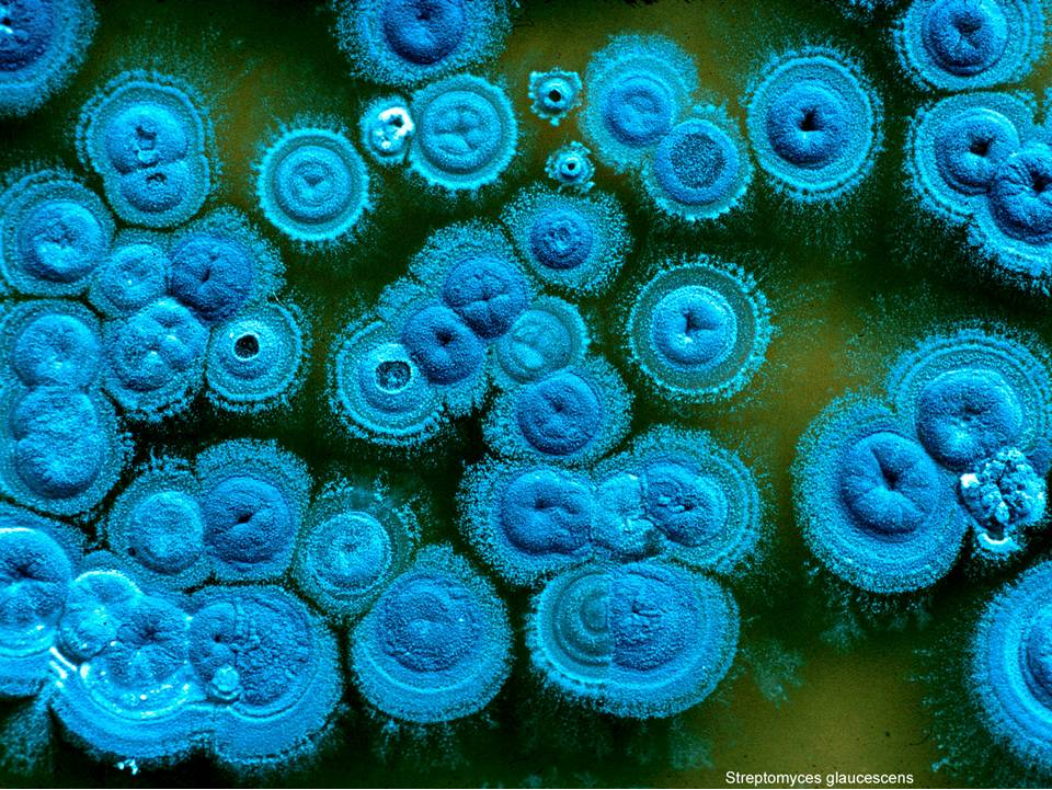
Image of Streptomyces glaucescens. Courtesy of Tobias Kieser, John Innes Centre
Mavericks of the Streptomyces family
Although these species are not considered to be important agents of infection, it is worth noting that not all Streptomyces bacteria are friends of humanity. Two rebels, Streptomyces somaliensis and Streptomyces sudanensis go against the grain by infecting people’s feet with actinomycotic mycetoma.
First described as ‘Madura foot’ in 1842, the disease is thought to date back to the Byzantine period and typically presents as cutaneous and subcutaneous tissue swelling, thickening, or painless nodule involving feet 80% of the time [6].
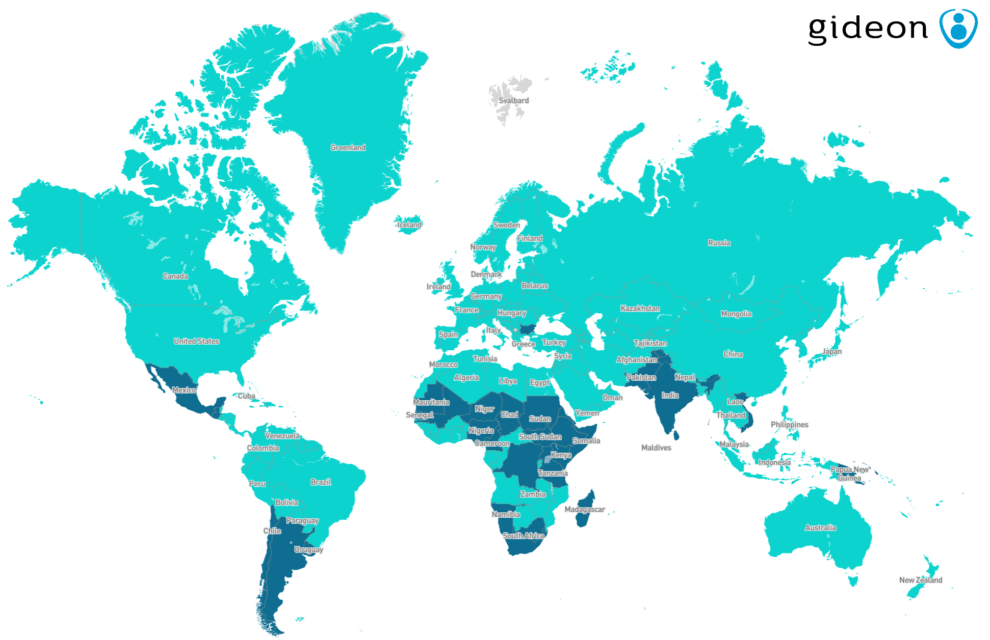
Mycetoma global distribution map, GIDEON. Dark blue color indicates recent reports of autochthonous cases.
Although bacterial mycetoma is endemic throughout the tropical world, S. somaliensis and S. sudanensis are only found in Eastern Africa, as names indicate. In other parts of the world, S.albus has been known to occasionally rear its head [7] in skin infections, although such occurrences are very rare.
And here’s the most fascinating fact – a distant ‘cousin’ of these pathogens, Streptomyces cattleya isan effective carbapenem used to treat Streptomyces spp. infections. A family feud in all its glory!
Why are bacteria named as fungi?
Streptomyces are about 450 million years old. Despite being a genus of bacteria, they are misleadingly suffixed with ‘-myces’, which stands for ‘fungi’ in Greek. This is because the first known example of the species contained branching filaments [8], a characteristic common to fungi.
Other actinobacteria, such as Mycobacteria and Actinomyces bear similar morphological features and thus carry a badge of mushroom in their names. Mycoplasmata – elusive gram-unidentifiable bacteria called ‘fungus form’, were named so by A. B. Frank in 1889. He thought ‘the “infection threads” of the organism were hyphae, and he knew of no hyphal-forming bacteria’ [9].
[1] H. Campbell, “Geosmin: Why We Like The Smell Of Air After A Storm”, American Council on Science and Health, 2018. [Online]. Available: https://www.acsh.org/news/2018/07/28/geosmin-why-we-smell-air-after-storm-13240. [Accessed: 11- Sep- 2020].
[2] I. BEAR and R. THOMAS, “Nature of Argillaceous Odour”, Nature, vol. 201, no. 4923, pp. 993-995, 1964. Available: 10.1038/201993a0 [Accessed 11 September 2020].
[3] K. Chater, “Streptomyces”, Brenner’s Encyclopedia of Genetics, pp. 565-567, 2013. Available: 10.1016/b978-0-12-374984-0.01483-2 [Accessed 11 September 2020].
[4] A. Hasani, A. Kariminik and K. Issazadeh, “Streptomycetes: Characteristics and Their Antimicrobial Activities”, International Journal of Advanced Biological and Biomedical Research, vol. 2, no. 1, pp. 63-75, 2014. Available: http://www.ijabbr.com/article_7033_7733c8235876d7ba635f6c831a916648.pdf. [Accessed 11 September 2020].
[6] V. Lichon and A. Khachemoune, “Mycetoma”, American Journal of Clinical Dermatology, vol. 7, no. 5, pp. 315-321, 2006. Available: 10.2165/00128071-200607050-00005 [Accessed 11 September 2020].
[7] M. Martin, A. Manteca, M. Castillo, F. Vazquez and F. Mendez, “Streptomyces albus Isolated from a Human Actinomycetoma and Characterized by Molecular Techniques”, Journal of Clinical Microbiology, vol. 42, no. 12, pp. 5957-5960, 2004. Available: 10.1128/jcm.42.12.5957-5960.2004 [Accessed 11 September 2020].
[8] K. Chater, “Streptomycesinside-out: a new perspective on the bacteria that provide us with antibiotics”, Philosophical Transactions of the Royal Society B: Biological Sciences, vol. 361, no. 1469, pp. 761-768, 2006. Available: 10.1098/rstb.2005.1758 [Accessed 12 September 2020].
[9] C. Krass and M. Gardner, “Etymology of the term Mycoplasma”, INTERNATIONAL JOURNAL of SYSTEMATIC BACTERIOLOGY, vol. 23, no. 1, pp. 62-64, 1973. Available: https://www.microbiologyresearch.org/docserver/fulltext/ijsem/23/1/ijs-23-1-62.pdf. [Accessed 12 September 2020].
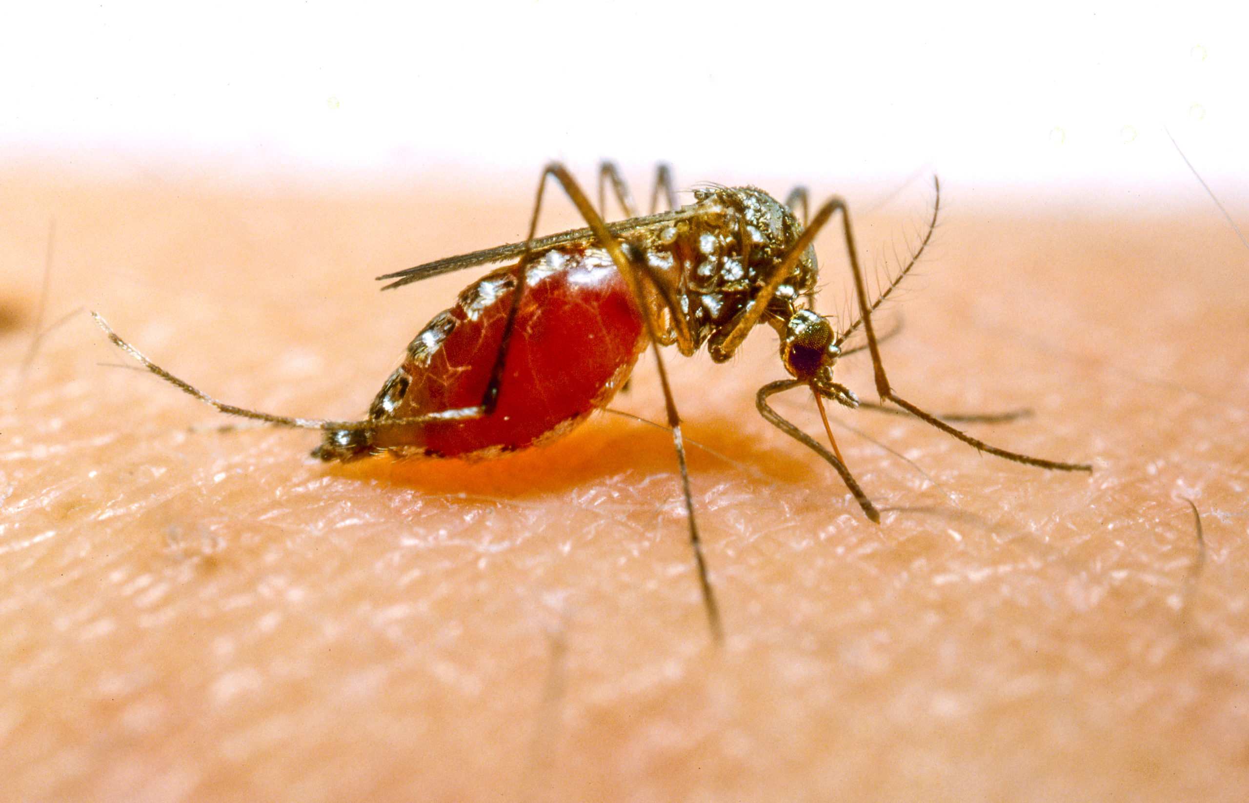
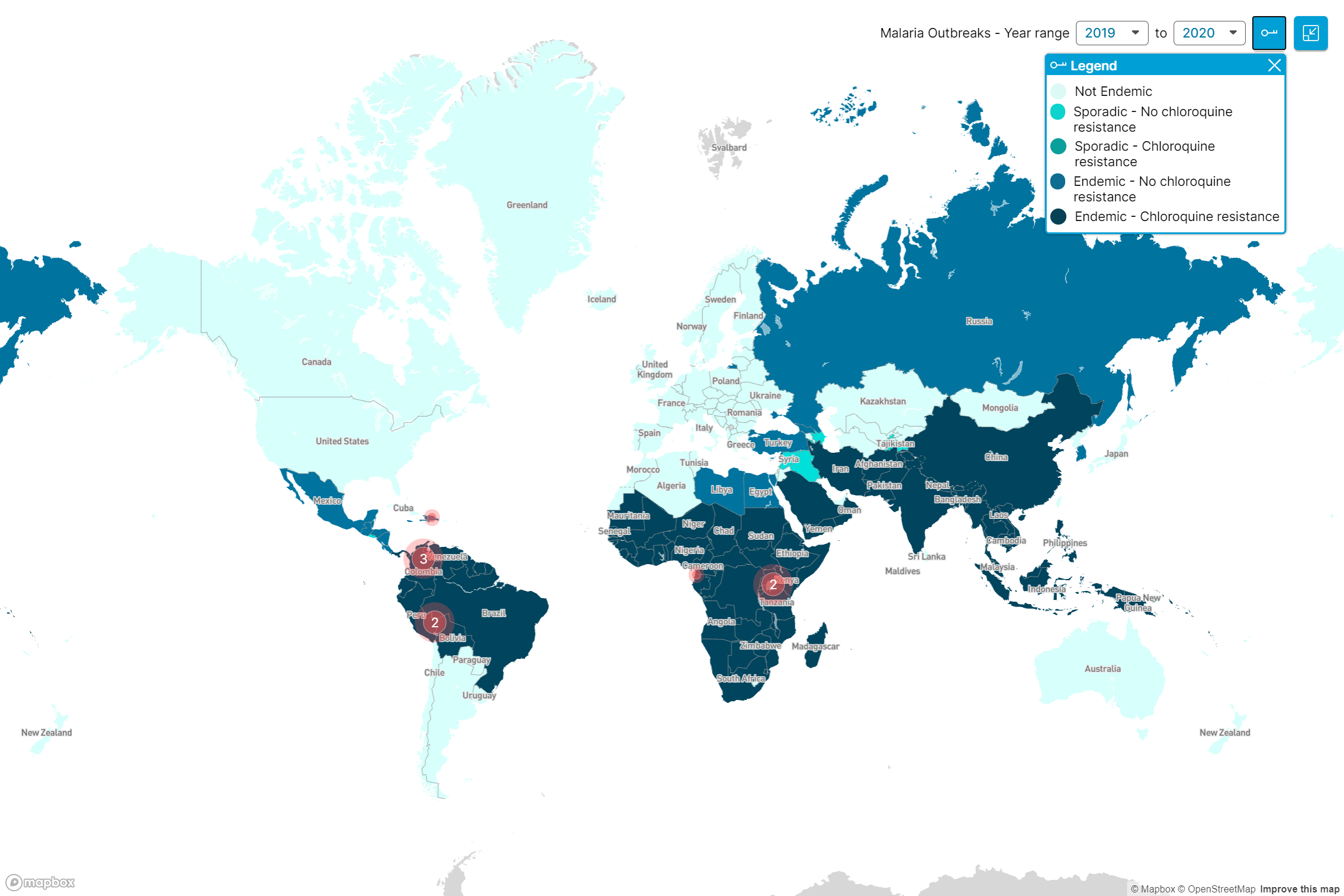
In one half of the world, the mosquito is seen to most as a minor annoyance, but for others, mosquitoes are synonymous with disease, pain, and death. Today is the World Mosquito Day and the perfect reminder of the devastating impact of such diseases as Malaria, Zika, and various kinds of Encephalitis for which mosquitoes are a major vector.
Malaria – a headline disease
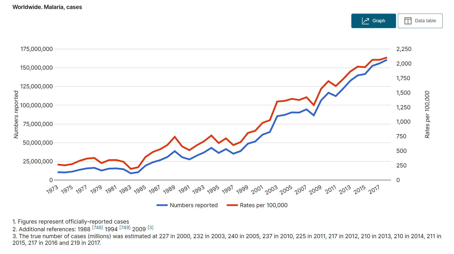
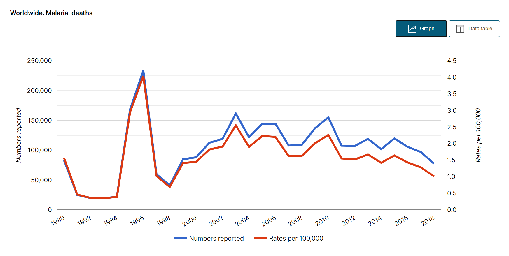
Malaria is the headline disease associated with mosquitoes and it was on this very day in 1897 that Sir Ronald Ross discovered that female mosquitoes can transmit malaria between humans. This was a major breakthrough in tackling the disease, yet despite significant progress, over 100 years on it is still the cause of tens of thousands of deaths every year, with annual case numbers on a steady incline.
‘Bad Air’ of the ancients
Malaria dates back thousands of years. Ancestral evidence found in 30-million-year-old amber shows that mosquitoes plagued humans from the earliest civilizations. It started having a significant impact on human survival roughly 10,000 years ago at the start of agriculture, and even Cleopatra is reputed to have slept under a mosquito net – though likely to avoid bites in general, rather than a preventative measure for the disease. Given the disease’s age, it is no surprise it has been referenced under many different names, such as Roman Fever and ‘Bad Air’ (Mal Aria), from which the modern name is derived.
A brief history of drugs
The most effective early drug was centered around Quinine, known since the 16th century and made from ground cinchona “fever” tree bark. Successfully synthesized in the early 20th century, it is the precursor to such drugs as Chloroquine. The revolutionary Methylene Blue – first synthetic antimalarial – was developed by Heinrich Caro. It helped differentiate between blood cells and the nuclei of malarial parasites. In a world without advanced microscopes, this was a significant breakthrough in identifying the disease.
Prevention is better than cure
The prevalence of the disease led to many efforts to try and prevent infection, but it wasn’t until Sir Ross proved the female mosquito as the vector in 1897 that targeted efforts could be made in limiting contact to mosquitoes, along with improving medicines. Nowadays, mosquito nets, insect repellents, and regular anti-insect medicines are commonplace in homes across the globe. The killing of mosquitoes was even cited as a selling point for the ill-fated insecticide DDT.
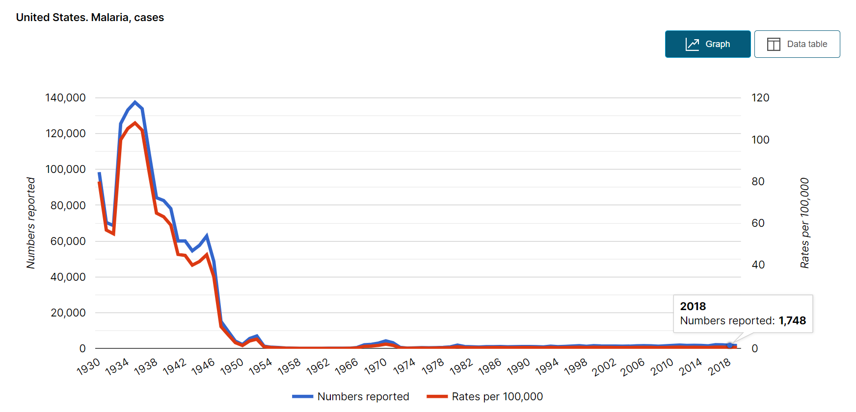
The United States was certified as “malaria-free” by the World Health Organisation in 1970, but even so over 1,000 cases are reported every year, virtually all imported from other countries.
While it is a preventable and curable disease, it is worth taking extra protection measures when traveling to high-risk areas, such as central Africa or India. A combination of symptoms such as headache, back pain, chills, sweating, myalgia, nausea, vomiting can be unpleasant, to say the least!
Did you like this article? Share it on social media!
Medical publishing has been significantly influenced by this year’s events. An unfortunate side effect of the COVID-19 pandemic has been an explosion of professional publications dealing with every possible aspect of the disease. Innumerable studies dissect the clinical features, epidemiology, imaging characteristics, and risk factors of a series of patients treated in hospitals or clinics.
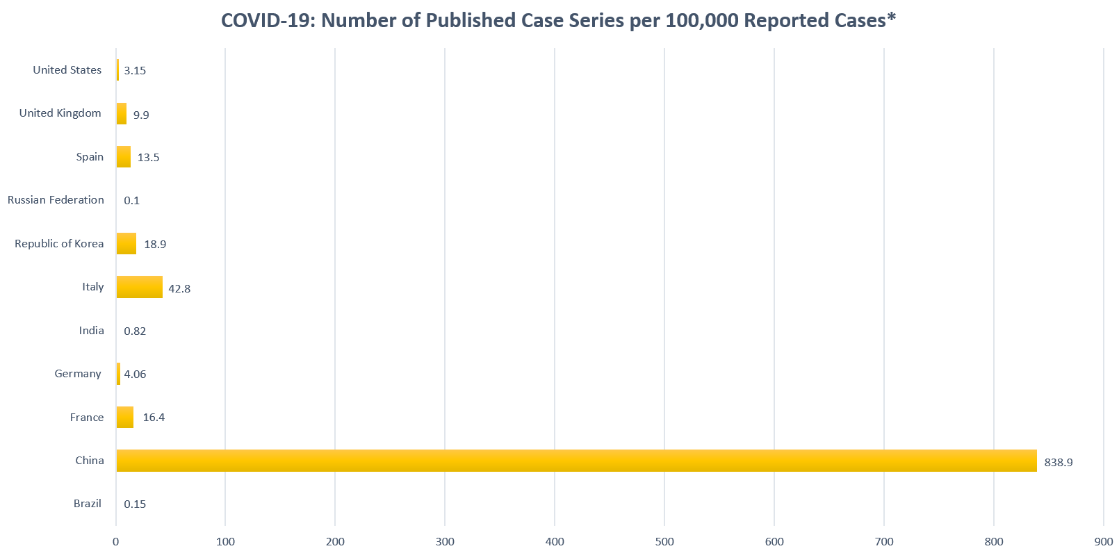
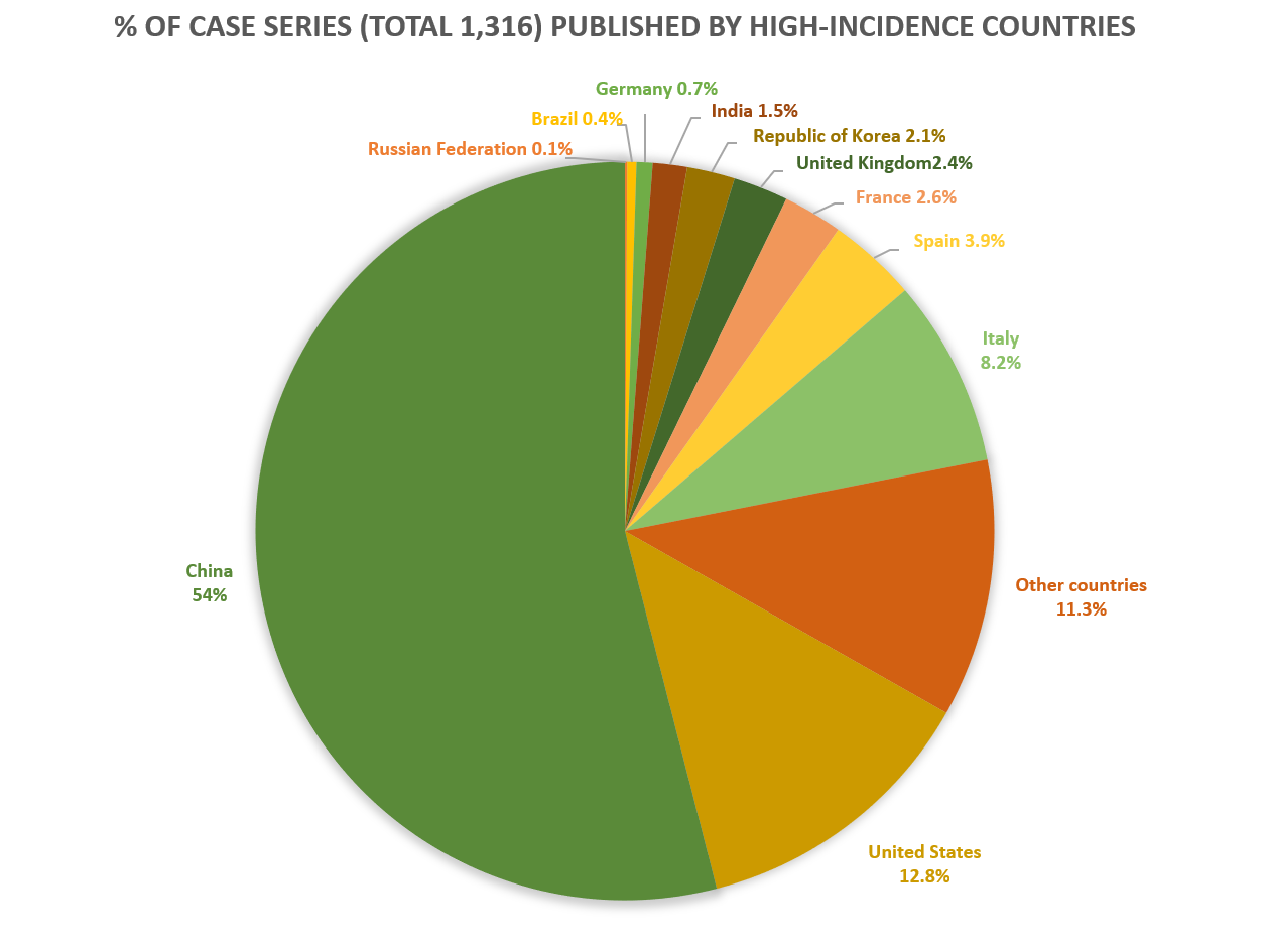
The GIDEON database collates all case series dealing with COVID-19 that have been cited in PubMed. These numbers do not include drug trials and disease prevalence surveys. 1,316 relevant series have been cited by PubMed as of August 13, published from 55 (28.2%) countries and dependencies which report the disease. Seven hundred and eleven series were published from China, encompassing 185,280 individual patients. The total number of COVID-19 cases reported by China to that date was 84,756. In other words, 2.19 Chinese patients appear in case series – for every 1 patient that is actually reported!
China alone accounts for 54.0% of all published case series, despite the fact that only 0.041% of the World’s cases were reported by that country. In part, these numbers might reflect the fact that “publishable data” were already accumulating in China before significant numbers were reported elsewhere, or delays that might be inherent in the publishing cycle itself. Other factors could include the relative numbers of clinical staff and institutions involved in research and publishing from any given country. Nevertheless, the unusually high number of case studies reported by China is difficult to explain. Assuming that “double-publication” can be discounted, large groups of patients are clearly “recycled” through multiple studies. From an epidemiological standpoint, this practice could actually contribute to the quality of data, since multiple studies of a given patient cohort can serve as excellent control groups for one another.
In the following bar graph, I’ve contrasted the ratio of published case series / reported cases among high-incidence countries. A pie graph contrasting the relative proportion of total case series from individual countries is also presented.
It is GIDEON’s mission to provide robust support to clinicians, microbiologists, and educators that interface with the infectious diseases field. As such, consulting with our users when developing the product is paramount to ensuring GIDEON continues to meet their expectations. On 12th August GIDEON closed its last alpha testing round, dedicated to gathering feedback on the brand new ‘Diagnose’ module. Medical experts from 26 countries have explored the new interface and shared their thoughts on what works and what can be improved.
WHAT IS ALPHA?
Alpha is an early release of a major product upgrade. Our first alpha testing round was focused on displaying our extensive database in a brand new ‘Explore’ module. The second one was geared towards clinicians who may use GIDEON in Point-of-Care settings to diagnose infectious diseases.
HOW DID IT GO?
The comments were overwhelmingly positive. Here are some of them:
“I just had a look and I am blown away both diagnosis modules. They are fantastic!”
“The product speaks for itself.”
“Having tried and tested each of the modules, GIDEON does a good job in making it quick and easy for a healthcare professional to access accurate and up to date information and that is the beauty and success of GIDEON.”
“The look and feel are great.”
“GIDEON remains an outstanding and easy to use, up to date resource. Commendable. Well done.”
We are so grateful for all the positivity as well as constructive criticism – it will make the product better for everyone.
WHAT’S NEXT?
A full beta release is currently planned for mid-September. It’s the same GIDEON data that you know and love, but like you’ve never seen before! The beta will include our brand new Lab module, designed to assist microbiologists. Check out a sneak-peek below:


 Medical publishing has been significantly influenced by this year’s events. An unfortunate side effect of the COVID-19 pandemic has been an explosion of professional publications dealing with every possible aspect of the disease. Innumerable studies dissect the clinical features, epidemiology, imaging characteristics, and risk factors of a series of patients treated in hospitals or clinics.